Skip to Content
MilliporeSigma
All Photos(2)

Documents

ABF210

Sigma-Aldrich

Anti-MDA5 Antibody

from rabbit, purified by affinity chromatography

Synonym(s):

MDA5 Antibody, Melanoma differentiation-associated protein 5, Interferon-induced with helicase C domain protein 1, IFIH1

Sign Into View Organizational & Contract Pricing


About This Item

UNSPSC Code:
12352203
eCl@ss:
32160702
NACRES:
NA.41

biological source

rabbit

Quality Level

antibody form

affinity isolated antibody

antibody product type

primary antibodies

clone

polyclonal

purified by

affinity chromatography

species reactivity

human

technique(s)

immunofluorescence: suitable
immunohistochemistry: suitable (paraffin)
western blot: suitable

UniProt accession no.

shipped in

wet ice

target post-translational modification

unmodified

Gene Information

human ... IFIH1(64135)

General description

Melanoma differentiation-associated protein 5 (MDA5), also known as Interferon-induced helicase C domain-containing protein 1 (IFIH1) is a member of the helicase family (RLR subfamily). MDA5 plays an important role in recognizing various viral components and triggers antiviral responses. When stimulated by dsRNA, MDA5 recruits adaptor protein VISA causing the activation of IRF-3 and NF-κB. MDA5 is widely expressed at low levels with barely detectable levels found in , testis and lung. Mutations in MDA5 have been associated with diabetes mellitus insulin-dependent type 19.

Specificity

This antibody recognizes the C-terminus of MDA5.

Immunogen

Epitope: C-terminus
KLH-conjugated linear peptide corresponding to the C-terminus of human MDA5.

Application

Detect Melanoma differentiation-associated protein 5 using this rabbit polyclonal antibody, Anti-MDA5 Antibody validated for use in western blotting, IHC (Paraffin) & Immunofluorescence.
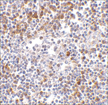
Immunohistochemistry Analysis: 5 µg/mL from a representative lot detected MDA5 in human lymph node tissue lysate.

Immunofluorescence Analysis: 20 μg/mL from a representative lot detected MDA5 in human lymph node cells.
Research Category
Inflammation & Immunology
Research Sub Category
Immunoglobulins & Immunology

Quality

Evaluated by Western Blotting in Daudi cell lysate.

Western Blotting Analysis: 2 µg/mL of this antibody detected MDA5 in 15 µg of Daudi cell lysate.

Target description

~117 kDa observed. Uncharacterized bands may appear in some lysates.

Linkage

Replaces: MABF198

Physical form

Antigen Affinity Purified
Purified rabbit polyclonal in buffer containing PBS with up to 0.1% sodium azide.

Storage and Stability

Stable for 1 year at 2-8°C from date of receipt.

Analysis Note

Control
Daudi cell Lysate

Other Notes

Concentration: Please refer to the Certificate of Analysis for the lot-specific concentration.

Disclaimer

Unless otherwise stated in our catalog or other company documentation accompanying the product(s), our products are intended for research use only and are not to be used for any other purpose, which includes but is not limited to, unauthorized commercial uses, in vitro diagnostic uses, ex vivo or in vivo therapeutic uses or any type of consumption or application to humans or animals.

Not finding the right product?  

Try our Product Selector Tool.

Storage Class Code

12 - Non Combustible Liquids

WGK

WGK 2

Flash Point(F)

Not applicable

Flash Point(C)

Not applicable


Certificates of Analysis (COA)

Search for Certificates of Analysis (COA) by entering the products Lot/Batch Number. Lot and Batch Numbers can be found on a product’s label following the words ‘Lot’ or ‘Batch’.

Already Own This Product?

Find documentation for the products that you have recently purchased in the Document Library.

Visit the Document Library

Lauren T Covert et al.
Rheumatology (Oxford, England), 63(1), 209-217 (2023-04-24)
To investigate pathogenic mechanisms underlying JDM, we defined the effect of type I IFN, IFN-α and IFN-β, on pediatric skeletal muscle function and expression of myositis-related proteins using an in vitro engineered human skeletal muscle model (myobundle). Primary myoblasts were
Ling Xu et al.
Proceedings of the National Academy of Sciences of the United States of America, 113(39), 10950-10955 (2016-09-14)
The function of the RIG-I-like receptors (RLRs; including RIG-I, MDA5, and LGP2) as key cytoplasmic sensors of viral pathogen-associated molecular patterns (PAMPs) has been subjected to numerous pathogenic challenges and has undergone a dynamic evolution. We found evolutionary evidence that

Our team of scientists has experience in all areas of research including Life Science, Material Science, Chemical Synthesis, Chromatography, Analytical and many others.

Contact Technical Service