Skip to Content
Merck
All Photos(2)

Documents

M7023

Sigma-Aldrich

Anti-Mouse IgG (whole molecule) antibody produced in rabbit

IgG fraction of antiserum, buffered aqueous solution

Sign Into View Organizational & Contract Pricing


About This Item

MDL number:
UNSPSC Code:
12352203
NACRES:
NA.46

biological source

rabbit

conjugate

unconjugated

antibody form

IgG fraction of antiserum

antibody product type

secondary antibodies

clone

polyclonal

form

buffered aqueous solution

technique(s)

indirect ELISA: 1:80,000
quantitative precipitin assay: 2.0 mg/mL

shipped in

dry ice

storage temp.

−20°C

target post-translational modification

unmodified

Looking for similar products? Visit Product Comparison Guide

Related Categories

General description

Immunoglobulin G (IgG) is a member of the immunoglobulin family and contains gamma (γ) heavy chain in the constant (C) region. Monomeric structure of IgG contains two identical heavy chains and two identical light chains with molecular weight of 50kDa and 25kDa respectively. The primary structure of this antibody also contains disulfide bonds implicated in linking the two heavy chains, linking the heavy and light chains and resides inside the chains. IgG is further subdivided into four classes namely, IgG1, IgG2, IgG3 and IgG4 with different heavy chains, named γ1, γ2, γ3 and γ4, respectively.

Specificity

The antiserum is determined to be immunospecific for mouse IgG by immunoelectrophoresis (IEP) versus normal mouse serum and mouse IgG. By Ouchterlony Double Diffusion (ODD) the product reacts with mouse IgG, mouse IgA and IgM.

Immunogen

Anti-Mouse IgG is produced in rabbit using purified mouse IgG as the immunogen. Whole antiserum is purified to provide an IgG fraction of antiserum.

Application

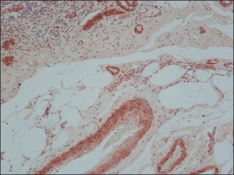
Anti-Mouse IgG (whole molecule) antibody produced in rabbit was used as secondary antibody in immunoblotting, immunoprecipitation and immunohistochemistry.

Biochem/physiol Actions

IgG antibody subtype is the most abundant of serum immunoglobulins of the immune system. It is secreted by B cells and is found in blood and extracellular fluids and provides protection from infections caused by bacteria, fungi and viruses. Maternal IgG is transferred to fetus through the placenta that is vital for immune defense of the neonate against infections.

Physical form

Solution in 0.01 M phosphate buffered saline pH 7.4, containing 15 mM sodium azide as preservative

Storage and Stability

For continuous use, store at 2-8 °C for up to one month. For extended storage, the solution may be frozen in working aliquots. Repeated freezing and thawing, or storage in "frost-free" freezers, is not recommended. If slight turbidity occurs upon prolonged storage, clarify the solution by centrifugation before use.

Disclaimer

Unless otherwise stated in our catalog or other company documentation accompanying the product(s), our products are intended for research use only and are not to be used for any other purpose, which includes but is not limited to, unauthorized commercial uses, in vitro diagnostic uses, ex vivo or in vivo therapeutic uses or any type of consumption or application to humans or animals.

Not finding the right product?  

Try our Product Selector Tool.

Storage Class Code

10 - Combustible liquids

WGK

WGK 1

Flash Point(F)

Not applicable

Flash Point(C)

Not applicable

Personal Protective Equipment

dust mask type N95 (US), Eyeshields, Gloves

Certificates of Analysis (COA)

Search for Certificates of Analysis (COA) by entering the products Lot/Batch Number. Lot and Batch Numbers can be found on a product’s label following the words ‘Lot’ or ‘Batch’.

Already Own This Product?

Find documentation for the products that you have recently purchased in the Document Library.

Visit the Document Library

A MacLaren et al.
Oncogene, 19(51), 5906-5918 (2000-12-29)
v-Jun shares the ability of the Myc, E1A, and E2F oncogenes to both sustain cell cycle progression and promote apoptosis in the absence of mitogenic stimulation. To gain an insight into the mechanism of apoptosis sensitization, we examined the possible
Lynsey Howard et al.
Stem cell research & therapy, 4(2), 36-36 (2013-04-27)
Differentiation of vascular endothelial cells (ECs) in clinically relevant numbers for injection into ischaemic areas could offer therapeutic potential in the treatment of cardiovascular conditions, including myocardial infarction, peripheral vascular disease and stroke. While we and others have demonstrated successful
Antibody structure, instability, and formulation.
Wang W, et al.
Journal of Pharmaceutical Sciences, 96(1), 1-26 (2007)
Shuobo Boboila et al.
Oncogene, 37(40), 5451-5465 (2018-06-09)
Despite the identification of MYCN amplification as an adverse prognostic marker in neuroblastoma, MYCN inhibitors have yet to be developed. Here, by integrating evidence from a whole-genome shRNA library screen and the computational inference of master regulator proteins, we identify
Seow Theng Ong et al.
The Journal of biological chemistry, 289(28), 19420-19434 (2014-05-30)
Rab GTPases control membrane traffic and receptor-mediated endocytosis. Within this context, Rab5a plays an important role in the spatial regulation of intracellular transport and signal transduction processes. Here, we report a previously uncharacterized role for Rab5a in the regulation of

Articles

Review the key factors that should figure in your decision to choose a secondary antibody. Learn about species, subclass, isotype, label, and more.

Review the key factors that should figure in your decision to choose a secondary antibody. Learn about species, subclass, isotype, label, and more.

Review the key factors that should figure in your decision to choose a secondary antibody. Learn about species, subclass, isotype, label, and more.

Review the key factors that should figure in your decision to choose a secondary antibody. Learn about species, subclass, isotype, label, and more.

Our team of scientists has experience in all areas of research including Life Science, Material Science, Chemical Synthesis, Chromatography, Analytical and many others.

Contact Technical Service