General description
Keratin, type II cytoskeletal 5 (UniProt P13647; also known as 58 kDa cytokeratin, CK-5, Cytokeratin-5, K5, Keratin-5, Type-II keratin Kb5) is encoded by the KRT5 (also known as DDD1, DM-EBS, EBSMCE, K-EBS, MP-EBS, WC-EBS) gene (Gene ID 3852) in human. Cytokeratin-5 (Keratin-5 or K5) belongs to a family of highly conserved epithelial-specific intermediate filament (IF) proteins. Each epithelium is characterized by its unique keratin expression pattern. In interfollicular epidermis, four major keratins are synthesized and co-polymerize in pairs, K5/K14 and K1/KI0, to form filaments. The expression of these two pairs is strictly regulated during normal keratinocyte differentiation. The K5/K14 filament is expressed in proliferating basal cells, while the K1/K10 filament is expressed during the transition to terminal differentiation and at the cessation of cell division. Similar to other IF members, Keratin-5 contains a central rod region composed of alpha helix segments (a.a. 168-203, 223-315, 339-477) that allow two different IF proteins, one type I (acidic) and one type II (basic) keratin, to intertwine into a coiled-coil formation via hydrophobic interaction. Kearain-5 is a type II keratin that pairs with the type I keratin-14 to form IF.
Specificity
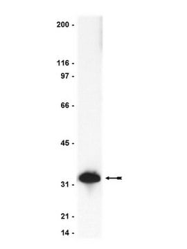
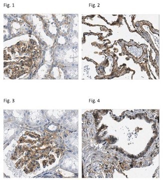
Clone D5/16B4 detected cytokeratin 5 by Western blotting using cytoskeletal preparations from human epidermis and non-cytokeratinizing epithelium (Abstract: European Symposium of the Biology of the Cytoskeleton, Helsinki, 18-21, 6, 89, 1989), as well as by staining tissue sections. Clone D5/16B4 also shows reactivity toward cytokeratin 6 and weak reactivity with cytokeratin 4 by Western blotting. No cross-reactivity to cytokeratin 1, 7, 8, 10, 13, 14, 18 or 19. Clone D5/16B4 immunostains basal cells and a part of the stratum spinosum in the normal pavement epithelium. Clone D5/16B4 is suitable for staining formalin-fixed tissue sections.
Immunogen
Purified cytokeratin 5 (Mischke, D., and Wild, G.A. (1987). J. Invest. Dermatol. 88(2):191-197; Wild, G.A., and Mischke, D. (1986). Exp. Cell Res. 162(1):114-126).
Application
Anti-Cytokeratin 5,6 Antibody, clone D5/16B4, Alexa Fluor™ 647 is an antibody against Cytokeratin 5,6 for use in Immunocytochemistry.
Research Category
Cell Structure
Research Sub Category
Cytoskeleton
The unconjugated antibody (Cat. No. MAB1620) is shown to be suitable also for immunofluorescence, immunohistochemistry, and Western blotting applications.
Quality
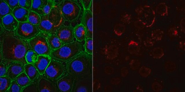
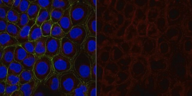
Evaluated by Immunocytochemistry in A431 cells.
Immunocytochemistry Analysis: A 1:100 dilution of this antibody detected Cytokeratin 5/6 in A431 cells.
Target description
Cytokeratin 4 (59 kDa), Cytokeratin 5 (58 kDa) and Cytokeratin 6 (56 kDa).
Physical form
Protein A purified
Purified mouse monoclonal IgG1 antibody conjugate in PBS with 15 mg/mL BSA and 0.1 % sodium azide.
Storage and Stability
Stable for 1 year at 2-8°C from date of receipt.
Other Notes
Concentration: Please refer to lot specific datasheet.
Legal Information
ALEXA FLUOR is a trademark of Life Technologies
Disclaimer
Unless otherwise stated in our catalog or other company documentation accompanying the product(s), our products are intended for research use only and are not to be used for any other purpose, which includes but is not limited to, unauthorized commercial uses, in vitro diagnostic uses, ex vivo or in vivo therapeutic uses or any type of consumption or application to humans or animals.


