Skip to Content
MilliporeSigma
All Photos(1)

Documents

IHCR2025-6

Sigma-Aldrich

IHC Select Anti-Cytokeratin AE1/AE3 (Pan cytokeratins) Antibody, prediluted, clone AE1/AE3

clone AE1/AE3, from mouse

Sign Into View Organizational & Contract Pricing


About This Item

UNSPSC Code:
12352203
eCl@ss:
32160702
NACRES:
NA.41

biological source

mouse

Quality Level

antibody form

purified antibody

antibody product type

primary antibodies

clone

AE1/AE3, monoclonal

species reactivity

human

manufacturer/tradename

Chemicon®
IHC Select

technique(s)

immunohistochemistry: suitable (paraffin)

isotype

IgG1

shipped in

wet ice

target post-translational modification

unmodified

Gene Information

human ... KRT1(3848)

General description

Keratins are a group of water-insoluble proteins that form monofilaments, a class of intermediate filament. These filaments form part of the cytoskeletal complex in epidermis and in most other epithelial tissues. Nineteen human epithelial keratins are resolved with two-dimensional gels electrophoresis (Moll, R., 1982). These can be divided into acid (pI <5.7) and basic (pI >6.0) subfamilies. The acidic keratins have molecular weights of 56.5, 55, 51, 50, 50′, 48, 46, 45, and 40 kD. The basic keratins have molecular weights of 65-67, 64, 59, 58, 56, and 52 kD. Members of the acidic and basic subfamilies are found together in pairs. The composition of keratin pairs varies with the epithelial cell type, stage of differentiation, cellular growth environment, and disease state (Sun, T.T., 1984; Cooper, D., 1985; Sun, T. T., 1985 ). The 56.5/65-67 kD pair is present in keratinized (differentiated) epidermis. The 55/64 kD pair is characteristic of normal (corneal-type) epithelial differentiation (Moll, R., 1982; Sun, T.T, 1984). The 51/59 kD pair is characteristic of the stratified squamous epithelial of internal organism such as esophagus and tongue (Moll, R., 1982; Cooper, D., 1985). The 51/58 kD pair is a keratinocyte marker; this pair is present in almost all stratified epithelia irrespective of the state of cellular stratification (Moll, R., 1982; Sun, T.T., 1984). The 48/56 kD pair is characteristic of hyper-proliferative (de-differentiated) keratinocytes (Moll, R., 1982; Weiss, R.A., 1984). The 45/52 kD pair and to a lesser extent, the 46/54 kD pair are characteristic of simple epithelia (Moll, R., 1982). The 40 kD keratin is present in most epithelia except adult epidermis (Moll, R., 1982).

Specificity

Anti-Keratin AE1 recognizes the 56.5, 50, 50′, 48, and 40 kD keratins of the acidic subfamily. Anti-keratin AE3 recognizes all members of the basic subfamily. AE1/AE3 reacts with both acidic and basic keratins. Staining is localized to the cytoplasm. AE1/AE3 specifically binds to antigens located in the cytoplasm of normal epithelia. Reacts with cells of epithelial origin including simple and stratified epithelia and epidermis.

Immunogen

Human epidermal keratin

application

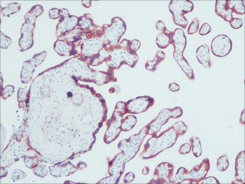
Antibody is prediluted and ready to use for Immunohistochemistry of formalin-fixed, paraffin-embedded tissues.

Pretreatment: Heat Induced Epitope Retrieval (HIER). Recommend Citrate Buffer, pH 6.0 (Cat. No. 21545). No signal was detected without Epitope retrieval.

Incubation: 10 minutes with IHC Select Detection Kits.

Cytokeratin AE1/AE3 has been prediluted for use as the primary antibody with Chemicon′s IHC Select Detection Kits and Protocols (Catalog Nos. DAB050, DET-HP1000, APR050, and DET-APR1000), but other supplier′s IHC detection systems may be used. For optimized protocol details, visit www.chemicon.com and select the protocols link under Cat. No.IHCR2025-6.
IHC Select Anti-Cytokeratin AE1/AE3 (Pan cytokeratins) Antibody, prediluted, clone AE1/AE3 is an antibody against Cytokeratin AE1/AE3 (Pan cytokeratins) for use in IH(P).
Research Category
Cell Structure
Research Sub Category
Cytokeratins

Physical form

Format: Purified
Purified monoclonal mouse antibody supplied in liquid format diluted in PBS, pH 7.2 with stabilizers, 0.2% Tween 20, and 0.1% Kathon as preservative.

Storage and Stability

Maintain at 2-8°C . Refer to vial for expiration dating.

Legal Information

CHEMICON is a registered trademark of Merck KGaA, Darmstadt, Germany

Disclaimer

Unless otherwise stated in our catalog or other company documentation accompanying the product(s), our products are intended for research use only and are not to be used for any other purpose, which includes but is not limited to, unauthorized commercial uses, in vitro diagnostic uses, ex vivo or in vivo therapeutic uses or any type of consumption or application to humans or animals.

Not finding the right product?  

Try our Product Selector Tool.

pictograms

Exclamation mark

signalword

Warning

hcodes

Hazard Classifications

Aquatic Chronic 3 - Skin Sens. 1

Storage Class

12 - Non Combustible Liquids

wgk_germany

WGK 2

flash_point_f

Not applicable

flash_point_c

Not applicable


Certificates of Analysis (COA)

Search for Certificates of Analysis (COA) by entering the products Lot/Batch Number. Lot and Batch Numbers can be found on a product’s label following the words ‘Lot’ or ‘Batch’.

Already Own This Product?

Find documentation for the products that you have recently purchased in the Document Library.

Visit the Document Library

Cindy Perez-Pacheco et al.
Clinical cancer research : an official journal of the American Association for Cancer Research, 29(13), 2501-2512 (2023-04-12)
Perineural invasion (PNI) in oral cavity squamous cell carcinoma (OSCC) is associated with poor survival. Because of the risk of recurrence, patients with PNI receive additional therapies after surgical resection. Mechanistic studies have shown that nerves in the tumor microenvironment
Jing Kong et al.
Cell communication & adhesion, 24(1), 11-18 (2018-05-08)
Salivary gland adenoid cystic carcinoma (SACC) is one of the most common malignancies in the oral and maxillofacial region. Carcinoma-associated fibroblast (CAF) is an important component in the tumor microenvironment and participates in SACC progression. In this study, we established
Ligia B Schmitd et al.
Neoplasia (New York, N.Y.), 20(7), 657-667 (2018-05-26)
A diagnosis of perineural invasion (PNI), defined as cancer within or surrounding at least 33% of the nerve, leads to selection of aggressive treatment in squamous cell carcinoma (SCC). Recent mechanistic studies show that cancer and nerves interact prior to
Christina Springstead Scanlon et al.
Nature communications, 6, 6885-6885 (2015-04-29)
Perineural invasion (PNI) is an indicator of poor survival in multiple cancers. Unfortunately, there is no targeted treatment for PNI since the molecular mechanisms are largely unknown. PNI is an active process, suggesting that cancer cells communicate with nerves. However

Our team of scientists has experience in all areas of research including Life Science, Material Science, Chemical Synthesis, Chromatography, Analytical and many others.

Contact Technical Service