Skip to Content
MilliporeSigma
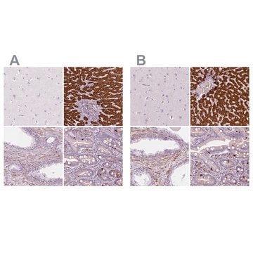
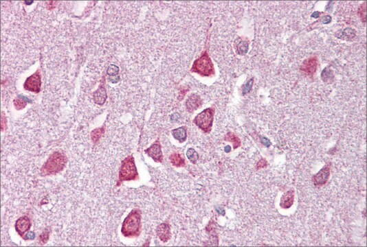
All Photos(1)

Documents

06-114

Sigma-Aldrich

Anti-Cathepsin D Antibody

from rabbit, purified by affinity chromatography

Synonym(s):

cathepsin D, cathepsin D (lysosomal aspartyl protease), lysosomal aspartyl peptidase, lysosomal aspartyl protease

Sign Into View Organizational & Contract Pricing


About This Item

UNSPSC Code:
12352203
eCl@ss:
32160702
NACRES:
NA.41

biological source

rabbit

antibody form

affinity isolated antibody

antibody product type

primary antibodies

clone

polyclonal

purified by

affinity chromatography

species reactivity

human

technique(s)

immunocytochemistry: suitable
western blot: suitable

NCBI accession no.

UniProt accession no.

shipped in

wet ice

target post-translational modification

unmodified

Gene Information

human ... CTSD(1509)

General description

Cathepsin D is synthesized as a 54kDa precursor which is proteolytically processed to an intermediate 48kDa single chain which matures into more stable 34kDa and 14kDa two chain form. It is an estrogen-regulated lysosomal protease that has been suggested to facilitate cancer cell migration and invasion by digesting the basement membrane, extracellular matrix, and connective tissue. Because of its mitogenic and proteolytic activities, it has been implicated as a prognostic marker in many tumor types, especially breast cancer. Cathepsin D is expressed in epithelial cells as well as in macrophages. Immunoreactivity of cancer cells and tissue infiltrating macrophages must be recorded separately.

Specificity

84% sequence homology to mouse and rat.
This antibody recognizes Cathepsin D.

Immunogen

Epitope: Heavy Chain
GST-tagged recombinant protein corresponding to human Cathepsin D at the heavy chain.

Application

Anti-Cathepsin D Antibody is an antibody against Cathepsin D for use in WB & IC.
Immunocytochemistry Analysis: 1:500 dilution from a previous lot detected Cathepsin D in A431 and HeLa cell lystes.
Research Category
Apoptosis & Cancer

Metabolism
Research Sub Category
Apoptosis - Additional

Quality

Evaluated by Western Blot in A431 cell lysate.

Western Blot Analysis: 1 µg/mL of this antibody detected Cathepsin D on 10 µg of A431 cell lysate.

Target description

Observed molecular weight at ~58 kDa Protein is glycosylated and may run slightly higher than predicted.
Predicted molecular weight of the active form is ~46 kDa and ~ 54 kDa for the precursor form.

Physical form

Affinity purified
Purified rabbit polyclonal in buffer containing 0.1 M Tris-Glycine (pH 7.4, 150 mM NaCl) with 0.05% sodium azide.

Storage and Stability

Stable for 1 year at 2-8°C from date of receipt.

Analysis Note

Control
A431 cell lysate

Other Notes

Concentration: Please refer to the Certificate of Analysis for the lot-specific concentration.

Disclaimer

Unless otherwise stated in our catalog or other company documentation accompanying the product(s), our products are intended for research use only and are not to be used for any other purpose, which includes but is not limited to, unauthorized commercial uses, in vitro diagnostic uses, ex vivo or in vivo therapeutic uses or any type of consumption or application to humans or animals.

Not finding the right product?  

Try our Product Selector Tool.

Storage Class

12 - Non Combustible Liquids

wgk_germany

WGK 1

flash_point_f

Not applicable

flash_point_c

Not applicable


Certificates of Analysis (COA)

Search for Certificates of Analysis (COA) by entering the products Lot/Batch Number. Lot and Batch Numbers can be found on a product’s label following the words ‘Lot’ or ‘Batch’.

Already Own This Product?

Find documentation for the products that you have recently purchased in the Document Library.

Visit the Document Library

Relationship among placental cadmium, lead, zinc, and copper levels in smoking pregnant women.
Turkan Kutlu,A Alev Karagozler,Engin M Gozukara
Biological trace element research null
Ganesh Vilas Shelke et al.
The Journal of cell biology, 222(6) (2023-05-22)
Exosomes are small vesicles that are secreted from cells to dispose of undegraded materials and mediate intercellular communication. A major source of exosomes is intraluminal vesicles within multivesicular endosomes that undergo exocytic fusion with the plasma membrane. An alternative fate

Our team of scientists has experience in all areas of research including Life Science, Material Science, Chemical Synthesis, Chromatography, Analytical and many others.

Contact Technical Service