Skip to Content
Merck
All Photos(1)

Documents

MAB3806

Sigma-Aldrich

Anti-ATM phosphoSer1981 Antibody, clone 10H11.E12

clone 10H11.E12, Chemicon®, from mouse

Synonym(s):

Ataxia Telangiectasia Mutated

Sign Into View Organizational & Contract Pricing


About This Item

UNSPSC Code:
12352203
eCl@ss:
32160702
NACRES:
NA.41

biological source

mouse

Quality Level

antibody form

purified immunoglobulin

antibody product type

primary antibodies

clone

10H11.E12, monoclonal

species reactivity

mouse, human

manufacturer/tradename

Chemicon®

technique(s)

immunocytochemistry: suitable
immunoprecipitation (IP): suitable
western blot: suitable

isotype

IgG1κ

NCBI accession no.

UniProt accession no.

shipped in

wet ice

target post-translational modification

phosphorylation (pSer1981)

Gene Information

human ... ATM(472)

General description

ATM is a 370 kDa nuclear phosphoprotein involved in the autosomal recessive disease Ataxia Telangiectasia (AT). Ataxia telangiectasia (AT) is a debilitating neurodegenerative disease that occurs early in childhood, resulting in ataxic movements and speech defects caused by cerebellar degeneration. The underlying cause of the disease is a biochemical dysfunction in the cellular response to specific types of DNA damage, correlated with mutations in the protein kinase ATM (Ataxia telangiectasia mutated). ATM not only is key in the signaling cascade that responds to DNA double-stranded breaks, but it also controls cell-cycle checkpoints and is therefore important in cancer prevention.

Specificity

Reacts with ATM Kinase phosphorylated at serine 1981. Clone 10H11.E12 is covered by US patent No. 6,916,627 and 7,108,992.

Immunogen

Epitope: phosphoSer1981
Synthetic peptide corresponding to amino acids 1974-1988 of human ATM (SLAFEEG[pS]QSTTISS).

Application

Anti-ATM Antibody, phosphoSer1981, clone 10H11.E12 is a Mouse Monoclonal Antibody for detection of ATM also known as Ataxia Telangiectasia Mutated & has been validated in IP, WB, ICC.
Research Category
Epigenetics & Nuclear Function
Research Sub Category
Cell Cycle, DNA Replication & Repair
Western blot: Detects a 370 kDa protein in crude extracts from irradiated human foreskin or mouse 3T3L1 cells.

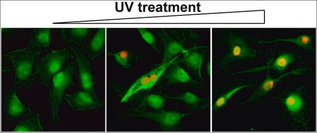
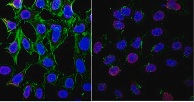
Immunocytochemistry: Foci are detected in irradiated human and mouse fibroblasts.

Immunoprecipitation: The antibody immunoprecipitates ATM from irradiated human and mouse cells.

Optimal working dilutions must be determined by end user.

Physical form

Format: Purified
Purified immunoglobulin presented as a liquid in 0.02M phosphate buffer containing 0.25M NaCl and 0.1% sodium azide.

Storage and Stability

Maintain at 2-8°C in undiluted aliquots for up to 6 months from date of receipt.


This antibody and certain aspects of its use are disclosed and claimed in pending U.S. Patent Applications published as U.S. Patent Publication Nos. 2003/0077661 and 2003/0157572.

Analysis Note

Control
Irradiated normal Human fibroblasts (no reactivity against non-irradiated cell extracts)

Other Notes

Concentration: Please refer to the Certificate of Analysis for the lot-specific concentration.

Legal Information

CHEMICON is a registered trademark of Merck KGaA, Darmstadt, Germany

Disclaimer

Unless otherwise stated in our catalog or other company documentation accompanying the product(s), our products are intended for research use only and are not to be used for any other purpose, which includes but is not limited to, unauthorized commercial uses, in vitro diagnostic uses, ex vivo or in vivo therapeutic uses or any type of consumption or application to humans or animals.

Not finding the right product?  

Try our Product Selector Tool.

Storage Class Code

10 - Combustible liquids

WGK

WGK 2

Flash Point(F)

Not applicable

Flash Point(C)

Not applicable


Certificates of Analysis (COA)

Search for Certificates of Analysis (COA) by entering the products Lot/Batch Number. Lot and Batch Numbers can be found on a product’s label following the words ‘Lot’ or ‘Batch’.

Already Own This Product?

Find documentation for the products that you have recently purchased in the Document Library.

Visit the Document Library

P Khongkow et al.
Oncogene, 33(32), 4144-4155 (2013-10-22)
FOXM1 is implicated in genotoxic drug resistance but its mechanism of action remains elusive. We show here that FOXM1-depletion can sensitize breast cancer cells and mouse embryonic fibroblasts (MEFs) into entering epirubicin-induced senescence, with the loss of long-term cell proliferation
Sarah Osterwald et al.
Journal of cell science, 128(10), 1887-1900 (2015-04-25)
The alternative lengthening of telomeres (ALT) mechanism allows cancer cells to escape senescence and apoptosis in the absence of active telomerase. A characteristic feature of this pathway is the assembly of ALT-associated promyelocytic leukemia (PML) nuclear bodies (APBs) at telomeres.
Nazmul Huda et al.
Cellular and molecular gastroenterology and hepatology, 14(2), 333-355 (2022-04-11)
Cellular senescence frequently is present in injured livers. The induction mechanism and the pathologic role are not always clear. We aimed to understand the dynamics of senescence induction and progression, and the mechanism responsible for the pathology using a mouse
Francesco Natale et al.
Nature communications, 8, 15760-15760 (2017-06-13)
Histone H2AX phosphorylation is an early signalling event triggered by DNA double-strand breaks (DSBs). To elucidate the elementary units of phospho-H2AX-labelled chromatin, we integrate super-resolution microscopy of phospho-H2AX during DNA repair in human cells with genome-wide sequencing analyses. Here we
Maxim Sorokin et al.
Oncotarget, 9(4), 5111-5124 (2018-02-13)
Acquired resistance to chemotherapy and radiation therapy is one of the major obstacles decreasing efficiency of treatment of the oncologic diseases. In this study, on the two cell lines (ovarian carcinoma SKOV-3 and neuroblastoma NGP-127), we modeled acquired resistance to

Our team of scientists has experience in all areas of research including Life Science, Material Science, Chemical Synthesis, Chromatography, Analytical and many others.

Contact Technical Service