MABN483
Anti-IRAP Antibody, Clone 3E1
clone 3E1, from mouse
同義詞:
Leucyl-cystinyl aminopeptidase, Cystinyl aminopeptidase, Insulin-regulated membrane aminopeptidase, Insulin-responsive aminopeptidase, IRAP, Oxytocinase, OTase, Placental leucine aminopeptidase, P-LAP, Leucyl-cystinyl aminopeptidase, pregnancy serum form
登入查看組織和合約定價
全部照片(1)
About This Item
分類程式碼代碼:
12352203
eCl@ss:
32160702
NACRES:
NA.41
推薦產品
生物源
mouse
品質等級
抗體表格
purified immunoglobulin
抗體產品種類
primary antibodies
無性繁殖
3E1, monoclonal
物種活性
human, mouse
技術
immunocytochemistry: suitable
western blot: suitable
同型
IgG1κ
NCBI登錄號
UniProt登錄號
運輸包裝
wet ice
目標翻譯後修改
unmodified
基因資訊
human ... LNPEP(4012)
一般說明
IRAP, also known as Leucyl-cystinyl aminopeptidase, Cystinyl aminopeptidase, Insulin-regulated membrane aminopeptidase, Insulin-responsive aminopeptidase (IRAP), Oxytocinase (OTase), or Placental leucine aminopeptidase (P-LAP) and encoded by the human gene name IRAP/OTASE, is an important aminopeptidase that plays an important role in maintaining homeostasis during pregnancy. IRAP degrades peptide hormones including oxytocin, vasopressin, and angiotensin III; it also appears to play a role in degrading neuronal peptides in the brain such as Met-enkephalin and dynorphin. IRAP also binds angiotensin IV and appears to be the receptor for angiotensin IV in the brain. IRAP is a membrane bound aminopeptidase and is often found within membrane vesicles that together with other proteins like GLUT4 translocates to the plasma membrane in response to insulin or oxytocin. A cleaved fragment of IRAP, the Leucyl-cystinyl aminopeptidase, is found in the blood during pregnancy, low in the first trimester but rising rapidly until birth, whereby it decreases rapidly, thus the secreted form is often used as a pregnancy marker. IRAP is highly expressed in placenta, heart, kidney and small intestine. IRAP is detected at lower levels in neuronal cells in the brain, in skeletal muscle, spleen, liver, testes and colon.
免疫原
Epitope: N-terminus
Recombinant protein corresponding to the N-terminus of human IRAP.
應用
Anti-IRAP, Clone 3E1 is an antibody against IRAP for use in WB, ICC.
Research Category
Neuroscience
Neuroscience
Research Sub Category
Developmental Signaling
Developmental Signaling
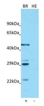
Western Blotting Analysis: A representative lot detected IRAP in 3T3-L1 cells treated with or without insulin (Garza, L.A., et al. (2000). JBC. 275(4):2560-2567).
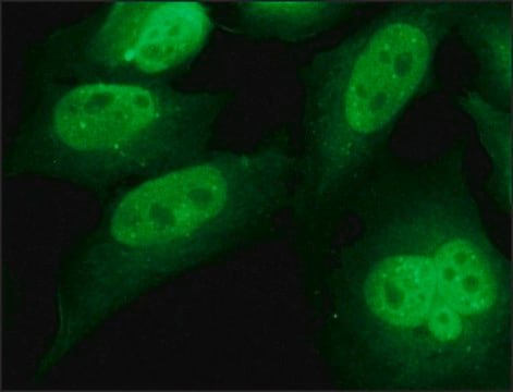
Immunocytochemistry Analysis: A representative lot detected IRAP in 3T3-L1 cells treated with or without insulin (Garza, L.A., et al. (2000). JBC. 275(4):2560-2567).
Immunocytochemistry Analysis: A representative lot detected IRAP in 3T3-L1 cells treated with or without insulin (Garza, L.A., et al. (2000). JBC. 275(4):2560-2567).
品質
Evaluated by Western Blotting in insulin treated NIH-3T3 cell lysate.
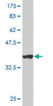
Western Blotting Analysis: 1.0 µg/mL of this antibody detected IRAP in 10 µg of insulin treated NIH-3T3 cell lysate.
Western Blotting Analysis: 1.0 µg/mL of this antibody detected IRAP in 10 µg of insulin treated NIH-3T3 cell lysate.
標靶描述
~150 kDa observed. The calculated molecular weight of this protein is 117 kDa, but can be observed at ~150 kDa due to high glycosylation. Uncharacterized band(s) may appear in some lysates.
外觀
Protein G Purified
Format: Purified
Purified mouse monoclonal IgG1κ in buffer containing 0.1 M Tris-Glycine (pH 7.4), 150 mM NaCl with 0.05% sodium azide.
儲存和穩定性
Stable for 1 year at 2-8°C from date of receipt.
其他說明
Concentration: Please refer to lot specific datasheet.
免責聲明
Unless otherwise stated in our catalog or other company documentation accompanying the product(s), our products are intended for research use only and are not to be used for any other purpose, which includes but is not limited to, unauthorized commercial uses, in vitro diagnostic uses, ex vivo or in vivo therapeutic uses or any type of consumption or application to humans or animals.
Not finding the right product?
Try our 產品選擇工具.
儲存類別代碼
12 - Non Combustible Liquids
水污染物質分類(WGK)
WGK 1
閃點(°F)
Not applicable
閃點(°C)
Not applicable
分析證明 (COA)
輸入產品批次/批號來搜索 分析證明 (COA)。在產品’s標籤上找到批次和批號,寫有 ‘Lot’或‘Batch’.。
Paula Nunes-Hasler et al.
Nature communications, 8(1), 1852-1852 (2017-11-28)
Antigen cross-presentation by dendritic cells (DC) stimulates cytotoxic T cell activation to promote immunity to intracellular pathogens, viruses and cancer. Phagocytosed antigens generate potent T cell responses, but the signalling and trafficking pathways regulating their cross-presentation are unclear. Here, we
我們的科學家團隊在所有研究領域都有豐富的經驗,包括生命科學、材料科學、化學合成、色譜、分析等.
聯絡技術服務