MABC94
Anti-Sphingosine 1-phosphate receptor 1 (S1P1) Antibody, clone 8B7.1
clone 8B7.1, from mouse
同義詞:
Sphingosine 1-phosphate receptor 1, S1P receptor 1, S1P1, Endothelial differentiation G-protein coupled receptor 1, Sphingosine 1-phosphate receptor Edg-1, S1P receptor Edg-1, CD363
登入查看組織和合約定價
全部照片(3)
About This Item
分類程式碼代碼:
12352203
eCl@ss:
32160702
NACRES:
NA.41
推薦產品
生物源
mouse
品質等級
抗體表格
purified immunoglobulin
抗體產品種類
primary antibodies
無性繁殖
8B7.1, monoclonal
物種活性
mouse, human, rat
技術
immunohistochemistry: suitable
western blot: suitable
同型
IgG2a
NCBI登錄號
UniProt登錄號
運輸包裝
wet ice
目標翻譯後修改
unmodified
基因資訊
human ... S1PR1(1901)
相關類別
一般說明
Sphingosine 1-phosphate receptor 1(S1PR1 or S1P1), otherwise known as EDG1, is a G-protein coupled receptor which is activated by lysosphingolipid sphingosine 1-phosphate (S1P) and lysophosphatidic acid (LPA). It is coupled to Gi-protein and MAP kinase signaling pathways, and may cooperate with other S1P receptors to regulate various cellular processes such as differentiation, actin remodeling, and migration. S1P1 is expressed in epithelial cells, fibroblasts, melanocytes, and smooth muscle cells.
免疫原
GST-tagged recombinant protein corresponding to mouse Sphingosine 1-phosphate receptor 1 (S1P1).
應用
Research Category
Apoptosis & Cancer
Apoptosis & Cancer
Research Sub Category
GPCR, cAMP/cGMP & Calcium Signaling
GPCR, cAMP/cGMP & Calcium Signaling
This Anti-Sphingosine 1-phosphate receptor 1 (S1P1) Antibody, clone 8B7.1 is validated for use in Western Blotting, IHC for the detection of Sphingosine 1-phosphate receptor 1 (S1P1).
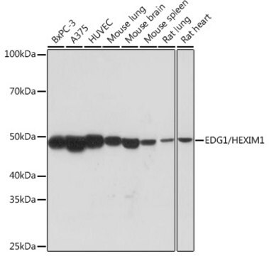
Western Blot Analysis: A 1:500 dilution from a representative lot detected Sphingosine 1-phosphate receptor 1 (S1P1) in human brain tissue lysate.
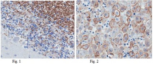
Immunohistochemistry Analysis: A 1:300 dilution from a representative lot detected Sphingosine 1-phosphate receptor 1 (S1P1) in rat choroid plexus tissue.
Immunohistochemistry Analysis: A 1:300 dilution from a representative lot detected Sphingosine 1-phosphate receptor 1 (S1P1) in rat choroid plexus tissue.
品質
Evaluated by Western Blot in mouse brain tissue lysate.
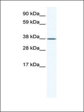
Western Blot Analysis: A 1:1,000 diluiton of this antibody detected Sphingosine 1-phosphate receptor 1 (S1P1) in 10 µg of mouse brain tissue lysate.
Western Blot Analysis: A 1:1,000 diluiton of this antibody detected Sphingosine 1-phosphate receptor 1 (S1P1) in 10 µg of mouse brain tissue lysate.
標靶描述
~40 kDa observed
外觀
Protein G Purified
Format: Purified
Purified mouse monoclonal IgG2a supernatant in buffer containing 0.1 M Tris-Glycine (pH 7.4), 150 mM NaCl with 0.05% sodium azide.
儲存和穩定性
Stable for 1 year at 2-8°C from date of receipt.
分析報告
Control
Mouse brain tissue lysate
Mouse brain tissue lysate
免責聲明
Unless otherwise stated in our catalog or other company documentation accompanying the product(s), our products are intended for research use only and are not to be used for any other purpose, which includes but is not limited to, unauthorized commercial uses, in vitro diagnostic uses, ex vivo or in vivo therapeutic uses or any type of consumption or application to humans or animals.
Not finding the right product?
Try our 產品選擇工具.
儲存類別代碼
12 - Non Combustible Liquids
水污染物質分類(WGK)
WGK 1
閃點(°F)
Not applicable
閃點(°C)
Not applicable
分析證明 (COA)
輸入產品批次/批號來搜索 分析證明 (COA)。在產品’s標籤上找到批次和批號,寫有 ‘Lot’或‘Batch’.。
Alhaji H Janneh et al.
Cell reports, 41(10), 111742-111742 (2022-12-09)
Crosstalk between metabolic and signaling events that induce tumor metastasis remains elusive. Here, we determine how oncogenic sphingosine 1-phosphate (S1P) metabolism induces intracellular C3 complement activation to enhance migration/metastasis. We demonstrate that increased S1P metabolism activates C3 complement processing through
Gustavo H Oliveira-Paula et al.
Science advances, 10(11), eadg9278-eadg9278 (2024-03-13)
Canonical Wnt and sphingosine-1-phosphate (S1P) signaling pathways are highly conserved systems that contribute to normal vertebrate development, with key consequences for immune, nervous, and cardiovascular system function; despite these functional overlaps, little is known about Wnt/β-catenin-S1P cross-talk. In the vascular
Jeffrey Thompson et al.
Aging and disease (2024-06-25)
Endothelial dysfunction and blood-brain barrier (BBB) leakage have been suggested as a fundamental role in the development of cerebral small vessel disease (SVD) pathology. However, the molecular and cellular mechanisms that link cerebral hypoxic hypoperfusion and BBB disruption remain elusive.
Sandeep K Singh et al.
Glia, 70(4), 712-727 (2021-12-28)
Astrocytes, the most abundant glial cells in the mammalian brain, directly associate with and regulate neuronal processes and synapses and are important regulators of brain development. Yet little is known of the molecular mechanisms that control the establishment of astrocyte
我們的科學家團隊在所有研究領域都有豐富的經驗,包括生命科學、材料科學、化學合成、色譜、分析等.
聯絡技術服務

