Skip to Content
Merck
All Photos(2)

Key Documents

MABS82

Sigma-Aldrich

Anti-phospho-p70 S6 Kinase (Thr389) Antibody, clone 10G7.1

clone 10G7.1, from mouse

Synonym(s):

Ribosomal protein S6 kinase beta-1, S6K-beta-1, S6K1, 70 kDa ribosomal protein S6 kinase 1, Short name=P70S6K1, p70-S6K 1, Ribosomal protein S6 kinase I, Serine/threonine-protein kinase 14A, p70 ribosomal S6 kinase alpha, p70 S6 kinase alpha, p70 S6K-alp

Sign Into View Organizational & Contract Pricing


About This Item

UNSPSC Code:
12352203
eCl@ss:
32160702
NACRES:
NA.41

biological source

mouse

Quality Level

antibody form

purified antibody

antibody product type

primary antibodies

clone

10G7.1, monoclonal

species reactivity

human, mouse

technique(s)

immunocytochemistry: suitable
western blot: suitable

isotype

IgG2aκ

NCBI accession no.

UniProt accession no.

shipped in

wet ice

target post-translational modification

phosphorylation (pThr389)

Gene Information

human ... RPS6KB1(6198)

General description

The p70 S6 kinase is a ubiquitous cytoplasmic protein that is activated in response to cytokines, and growth factors including the epidermal growth factor and fibroblast growth factor. It lies downstream of the mTOR/PI3K pathway and is phosphorylated on multiple residues: Thr389, Thr229, Thr421, Ser371, Ser404, Ser411, Ser418, and Ser424. Thr389, Ser371, and Ser404 are located in the linker domain; whereas Thr421, Ser411, Ser418, Ser424 are located in the autoinhibitory domain, and Thr229 is found in the catalytic domain. These sites may be phosphorylated in a hierarchical sequence leading to the activation of the p70 S6 kinase. The p70 S6 kinase phosphorylates the S6 protein on the 40S ribosomal protein (rpS6), and plays a role in multiple processes including regulation of mRNA molecules with 5’-terminal oligopyrimidine tracts, protein synthesis, cell growth, proliferation, and cell survival. The activity of this kinase may be important in diabetes, cancer, and aging.

Specificity

This antibody recognizes p70 S6 Kinase phosphorylated at (Thr389). This antibody also recognizes isoforms Alpha I and Alpha II.

Immunogen

Epitope: Phosphorylated (Thr389)
KLH-conjugated linear peptide corresponding to mouse p70 S6 Kinase phosphorylated at (Thr389).

Application

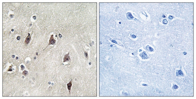
Immunocytochemistry Analysis: A 1:500 dilution from a representative lot detected p70 S6 Kinase in A431 cells.
Research Category
Signaling
Research Sub Category
Cell Cycle, DNA Replication & Repair
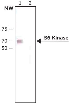
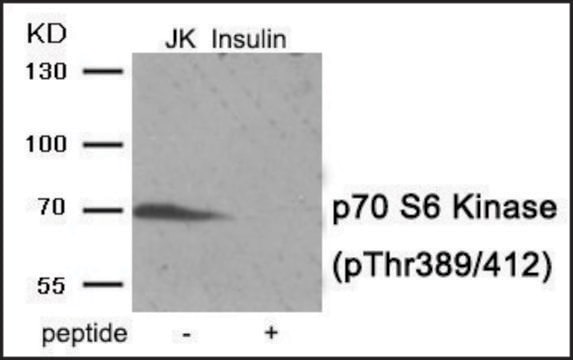
This Anti-phospho-p70 S6 Kinase (Thr389) Antibody, clone 10G7.1 is validated for use in Western Blotting, ICC for the detection of phospho-p70 S6 Kinase (Thr389).

Quality

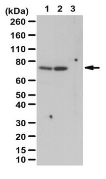
Evaluated by Western Blot in untreated and serum treated NIH/3T3 cell lysates.

Western Blot Analysis: 1 µg/mL of this antibody detected p70 S6 Kinase on 10 µg of untreated and serum treated NIH/3T3 cell lysates.

Target description

~70/85 kDa observed. This protein recognizes isoforms Alpha I and Alpha II. Uncharacterized bands appear at ~18 and 200 kDa in some lysates depending on treatment conditions.

Linkage

Replaces: 04-392

Physical form

Format: Purified
Protein G Purified
Purified mouse monoclonal IgG2aκ in buffer containing 0.1 M Tris-Glycine (pH 7.4), 150 mM NaCl with 0.05% sodium azide.

Storage and Stability

Stable for 1 year at 2-8°C from date of receipt.

Analysis Note

Control
Untreated and serum treated NIH/3T3 cell lysates

Other Notes

Concentration: Please refer to the Certificate of Analysis for the lot-specific concentration.

Disclaimer

Unless otherwise stated in our catalog or other company documentation accompanying the product(s), our products are intended for research use only and are not to be used for any other purpose, which includes but is not limited to, unauthorized commercial uses, in vitro diagnostic uses, ex vivo or in vivo therapeutic uses or any type of consumption or application to humans or animals.

Not finding the right product?  

Try our Product Selector Tool.

Storage Class Code

12 - Non Combustible Liquids

WGK

WGK 1

Flash Point(F)

Not applicable

Flash Point(C)

Not applicable


Certificates of Analysis (COA)

Search for Certificates of Analysis (COA) by entering the products Lot/Batch Number. Lot and Batch Numbers can be found on a product’s label following the words ‘Lot’ or ‘Batch’.

Already Own This Product?

Find documentation for the products that you have recently purchased in the Document Library.

Visit the Document Library

Darren M Blackburn et al.
iScience, 27(7), 110241-110241 (2024-07-17)
Adult stem cells play a critical role in tissue repair and maintenance. In tissues with slow turnover, including skeletal muscle, these cells are maintained in a mitotically quiescent state yet remain poised to re-enter the cell cycle to replenish themselves
Yu-Ting Kang et al.
Oncotarget, 8(62), 105536-105552 (2017-12-30)
Autophagy is an intracellular recycling and degradation process for regulating tumor progression, survival and drug resistance. Nickel compounds have been identified as human carcinogens. However, the role of nickel-induced autophagy in lung carcinogenesis has not yet been fully elucidated. In
Hui-Ping Hsu et al.
Cancer medicine, 12(2), 1588-1601 (2022-06-28)
Tumor cells may aberrantly express metabolic enzymes to adapt to their environment for survival and growth. Targeting cancer-specific metabolic enzymes is a potential therapeutic strategy. Phosphoenolpyruvate carboxykinase (PEPCK) catalyzes the conversion of oxaloacetate to phosphoenolpyruvate and links the tricarboxylic acid
Yasushi Nakai et al.
Oncology reports, 37(1), 227-234 (2016-11-15)
We examined extracellular signal‑regulated kinase (ERK), 4E‑binding protein 1 (4EBP1) and p70 ribosomal S6 kinase (p70) as potential biomarkers for pretreatment prediction of the prognosis of patients with metastatic renal cell carcinoma (RCC) treated with sorafenib, sunitinib or everolimus. 786‑O and 769‑P
Debasish Boral et al.
Cancers, 12(6) (2020-06-25)
Despite widespread knowledge that bone marrow-resident breast cancer cells (BMRCs) affect tumor progression, signaling mechanisms of BMRCs implicated in maintaining long-term dormancy have not been characterized. To overcome these hurdles, we developed a new experimental model of clinical dormancy employing

Our team of scientists has experience in all areas of research including Life Science, Material Science, Chemical Synthesis, Chromatography, Analytical and many others.

Contact Technical Service