830-05A
Human Mammary Epithelial Cells: HMEpC, adult
Synonym(s):
HMEpc cells
Sign Into View Organizational & Contract Pricing
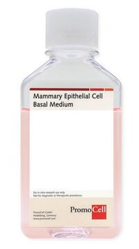
All Photos(2)
About This Item
UNSPSC Code:
41106514
NACRES:
NA.81
Recommended Products
biological source
human mammary glands (normal)
Quality Level
packaging
pkg of 500,000 cells
manufacturer/tradename
Cell Applications, Inc
growth mode
Adherent
karyotype
2n = 46
morphology
epithelial
technique(s)
cell culture | mammalian: suitable
relevant disease(s)
cancer
shipped in
liquid nitrogen
storage temp.
−196°C
General description
Lot specific orders are not able to be placed through the web. Contact your local sales rep for more details.
Mammary Epithelial Cells provide an excellent model system to study many aspects of epithelial function and disease, particularly those related to cancerogenesis.
HMEpC have been utilized in numerous research publications, for example to:
HMEpC, along with Human Prostate Epithelial Cells, have also been used in a study demonstrating that TGF-b1 induces Smad 1/5/8 and Smad 2/3 phosphorylation and BMP signaling results only in Smad 1/5/8 phosphorylation in these primary epithelial cells, while in cancer cells BMP also elicits Smad2/3 activation, contributing to cancerogenesis (Holtzhausen, 2013).
Characterization: the cells have a characteristic morphology consistent with an epithelial origin and are positive for epithelial cell marker cytokeratin 18.
Mammary Epithelial Cells provide an excellent model system to study many aspects of epithelial function and disease, particularly those related to cancerogenesis.
HMEpC have been utilized in numerous research publications, for example to:
- Investigate the role of exosomes secreted by cancer cells in formation of tumor permissive microenvironment through manipulation of normal mammary epithelium (Dutta, 2014)
- Serve as control in a study investigating antitumor properties of cannabinoids (Ligresti, 2006) and stem cell microenvironment (Rostovit, 2008)
- Determine that differential expression of glycoproteins allows to classify human breast cells into normal, benign, malignant, basal, and luminal groups (Yen, 2011, 2013; Timpe, 2013)
- Identify ALDH isoform 5A1 as a potential target for treatment of human breast ductal carcinoma (Kaur, 2012); and determine that combination of an anti-EGFR anti-VEGFR treatment using ZD6474 with phototherapy (UV-B) is more effective in treating breast cancer than either treatment alone (Sarkar, 2013)
- Demonstrate that although SIRT deacetylates p53, it does not play a role in cell survival following DNA damage (Solomon, 2006)
- Demonstrate the important roles of tumor suppressor Maspin by showing that it promotes mammary epithelial differentiation via its interaction with IRF6 (Bailey, 2008); mediates effects of IFN-γ on vacuolar pH, cathepsin D processing and autophagy, protects extracellular matrix (ECM) from degradation in normal mammary epithelia, and that Maspin loss in metastatic cancer leads to unrestricted ECM degradation, contributing to metastasis (Khalkhali-Ellis, 2007, 2008); and show that loss of EcSOD expression also promotes invasiveness by disrupting ECM (Teoh-Fitzgerald, 2012, 2013)
- Investigate the role of shortened telomeres in initiation of genomic instability, cytokinesis failure and polyploidy (Tusell, 2008; Soler, 2009; Pampalona, 2012); and elucidate the role of Myc in malignancy by studying its ability to transform primary epithelial cells (Thibodeaux, 2009)
- Demonstrate, along with Human dermal Fibroblasts, that resveratrol inhibits mono-ubiquitination of histone H2B (Gao, 2011)
HMEpC, along with Human Prostate Epithelial Cells, have also been used in a study demonstrating that TGF-b1 induces Smad 1/5/8 and Smad 2/3 phosphorylation and BMP signaling results only in Smad 1/5/8 phosphorylation in these primary epithelial cells, while in cancer cells BMP also elicits Smad2/3 activation, contributing to cancerogenesis (Holtzhausen, 2013).
Characterization: the cells have a characteristic morphology consistent with an epithelial origin and are positive for epithelial cell marker cytokeratin 18.
Cell Line Origin
Mammary Gland
Application
antitumor properties, glycoprotein expression, breast cancer treatment approaches, DNA damage, cell differentiation, effects of growth factors
Components
Cell Basal Medium containing 10% FBS & 10% DMSO
Preparation Note
- 5th passage, >500,000 cells in Cell Basal Medium containing 10% FBS & 10% DMSO
- Can be cultured at least 16 doublings
Subculture Routine
Please refer to the HMEpC Culture Protocol.
Disclaimer
RESEARCH USE ONLY. This product is regulated in France when intended to be used for scientific purposes, including for import and export activities (Article L 1211-1 paragraph 2 of the Public Health Code). The purchaser (i.e. enduser) is required to obtain an import authorization from the France Ministry of Research referred in the Article L1245-5-1 II. of Public Health Code. By ordering this product, you are confirming that you have obtained the proper import authorization.
Storage Class Code
11 - Combustible Solids
WGK
WGK 3
Flash Point(F)
Not applicable
Flash Point(C)
Not applicable
Certificates of Analysis (COA)
Search for Certificates of Analysis (COA) by entering the products Lot/Batch Number. Lot and Batch Numbers can be found on a product’s label following the words ‘Lot’ or ‘Batch’.
Already Own This Product?
Find documentation for the products that you have recently purchased in the Document Library.
Customers Also Viewed
Karthic Chandran et al.
Oncotarget, 7(13), 15577-15599 (2015-12-02)
Inflammatory and invasive breast cancers are aggressive and require better understanding for the development of new treatments and more accurate prognosis. Here, we detected high expression of PPARα in human primary inflammatory (SUM149PT) and highly invasive (SUM1315MO2) breast cancer cells
Our team of scientists has experience in all areas of research including Life Science, Material Science, Chemical Synthesis, Chromatography, Analytical and many others.
Contact Technical Service


