
推荐产品


生物来源
mouse
质量水平
重组
expressed in HEK 293 cells
偶联物
unconjugated
抗体形式
purified antibody
抗体产品类型
primary antibodies
克隆
P1B5, recombinant monoclonal
描述
recombinant, expressed in HEK 293 cells
产品线
ZooMAb® learn more
表单
lyophilized
分子量
calculated mol wt 117 kDa
纯化方式
using protein G
种属反应性
human
包装
antibody small pack of 25 μL
环保替代产品特性
Waste Prevention
Designing Safer Chemicals
Design for Energy Efficiency
Learn more about the Principles of Green Chemistry.
增强验证
recombinant expression
Learn more about Antibody Enhanced Validation
sustainability
Greener Alternative Product
技术
ELISA: suitable
affinity binding assay: suitable
flow cytometry: suitable
immunocytochemistry: suitable
同位素/亚型
IgG1κ
表位序列
Extracellular domain
Protein ID登记号
UniProt登记号
环保替代产品分类
运输
ambient
储存温度
2-8°C
相关类别
一般描述
We are committed to bringing you greener alternative products, which adhere to one or more of The 12 Principles of Green Chemistry.This antibody is Preservative-free, produced without the harm or sacrifice of animals and exceptionally stable to allow for ambient shipping and storage if needed and thus aligns with "Waste Prevention", "Designing Safer Chemicals" and "Design for Energy Efficiency". Click here for more information.
ZooMAb® antibodies represent an entirely new generation of recombinant monoclonal antibodies. Each ZooMAb® antibody is manufactured using our proprietary recombinant expression system, purified to homogeneity, and precisely dispensed to produce robust and highly reproducible lot-to-lot consistency. Only top-performing clones are released for use by researchers. Each antibody is validated for high specificity and affinity across multiple applications, including its most commonly used application. ZooMAb® antibodies are reliably available and ready to ship when you need them.
特异性
Clone P1B5 is a ZooMAb® mouse recombinant monoclonal antibody that detects Integrin 3 (ITGA3). It targets an epitope within the extracellular domain.
免疫原
HT1080 cells.
应用
Quality Control Testing
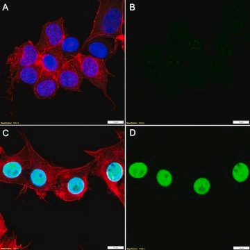
Evaluated by Immunocytochemistry in A431 cells.
Immunocytochemistry Analysis: A 1:1,000 dilution of this antibody detected ITGA3 (Integrin 3) in A431 cells.
Tested Applications
Flow Cytometry Analysis: 0.1 µg from a representative lot detected ITGA3 (Integrin 3) in one million HT1080 cells.
Affinity Binding Assay: A representative lot of this antibody bound recombinant fragment corresponding to the extracellular domain of Human ITGA3 (Integrin 3) with a KD of 7.3 x 10⁻⁶ in an affinity binding assay.
Enzyme Immunoassay (ELISA) Analysis: A serial of dilutions from a representative lot detected r ecombinant fragment corresponding to the extracellular domain of Human ITGA3 (Integrin 3) recombinant fragment.
Note: Actual optimal working dilutions must be determined by end user as specimens, and experimental conditions may vary with the end user.
目标描述
Integrin alpha-3 (UniProt: P26006; also known as CD49 antigen-like family member C, FRP-2, Galactoprotein B3, GAPB3, VLA-3 subunit alpha, CD49c) is encoded by the ITGA3 (also known as MSK18) gene (Gene ID: 3675) in human. Integrins are heterodimeric integral membrane proteins composed of an α subunit and β subunit that function in cell surface adhesion and signaling. They contain a large extracellular domain responsible for ligand binding, a single transmembrane domain, and a cytoplasmic domain. The exact combination of various α- and β-subunits dictates the binding specificity of integrins to different ECM components. Although both subunits are required for adhesion, the binding specificity primarily depends on the extracellular portion of the α-subunit. The structural and functional diversity of the integrin family is based upon the pairing abilities of the individual α and β subunits. Integrin α3 is a single-pass type I membrane glycoprotein that is synthesized with a signal peptide (aa 1-32), which is subsequently cleaved off to generate the mature form that contains an extracellular domain (aa 33-991), a transmembrane domain (aa 992-1014), and a cytoplasmic domain (aa 1015-1051). It has two chains- a heavy chain (aa 33-872) and a light chain (aa 876-1051). Integrin α3 dimerizes with integrin β1 to form a receptor for fibronectin, laminin, collagen, epiligrin, thrombospondin, and chondroitin sulfate proteoglycan 4 (CSPG4). It provides a docking site for FAP (seprase) at invadopodia plasma membranes in a collagen-dependent manner and hence may participate in the adhesion, formation of invadopodia and matrix degradation processes, promoting cell invasion. Overexpression of ITGA3 has been reported in intrahepatic cholangiocarcinoma. It promotes proliferation and cell cycle progression leading to poor prognosis. This ZooMAb® recombinant monoclonal antibody, generated by our propriety technology, offers significantly enhanced specificity, Affinity™, reproducibility, and stability over conventional monoclonals. (Ref.: Huang, Y., et al. (2018). Biomed. Res. Int. 2018; Article 2352139; Mueller, SC., et al. (1999). J. Biol. Chem. 274(35); 24947-24952).
外形
Purified recombinant mouse monoclonal antibody IgG, lyophilized in PBS, 5% Trehalose, normal appearance a coarse or translucent resin. The PBS/trehalose components in the ZooMAb formulation can have the appearance of a semi-solid (bead like gel) after lyophilization. This is a normal phenomenon. Please follow the recommended reconstitution procedure in the data sheet to dissolve the semi-solid, bead-like, gel-appearing material. The resulting antibody solution is completely stable and functional as proven by full functional testing. Contains no biocide or preservatives, such as azide, or any animal by-products. Larger pack sizes provided as multiples of 25 µL.
储存及稳定性
Recommend storage of lyophilized product at 2-8°C; Before reconstitution, micro-centrifuge vials briefly to spin down material to bottom of the vial; Reconstitute each vial by adding 25 µL of filtered lab grade water or PBS; Reconstituted antibodies can be stored at 2-8°C, or -20°C for long term storage. Avoid repeated freeze-thaws.
法律信息
Affinity is a trademark of Mine Safety Appliances Co.
ZooMAb is a registered trademark of Merck KGaA, Darmstadt, Germany
免责声明
Unless otherwise stated in our catalog or other company documentation accompanying the product(s), our products are intended for research use only and are not to be used for any other purpose, which includes but is not limited to, unauthorized commercial uses, in vitro diagnostic uses, ex vivo or in vivo therapeutic uses or any type of consumption or application to humans or animals.
未找到合适的产品?
试试我们的产品选型工具.
储存分类代码
11 - Combustible Solids
WGK
WGK 1
闪点(°F)
Not applicable
闪点(°C)
Not applicable
我们的科学家团队拥有各种研究领域经验,包括生命科学、材料科学、化学合成、色谱、分析及许多其他领域.
联系技术服务部门