
$256.00
预计发货时间2025年5月13日详情
新价格,新优惠!
推荐产品


生物来源
mouse
抗体形式
purified from hybridoma cell culture
抗体产品类型
primary antibodies
克隆
COL-10, monoclonal
表单
buffered aqueous solution
分子量
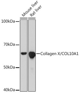
~60 kDa
种属反应性
deer, porcine, human
包装
antibody small pack of 25 μL
浓度
~1 mg/mL
技术
immunoblotting: suitable
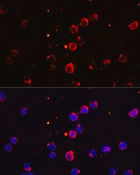
immunofluorescence: 5-10 μg/mL using human osteosarcoma SaOS-2 cells
immunohistochemistry: suitable
同位素/亚型
IgM
UniProt登记号
运输
dry ice
储存温度
−20°C
靶向翻译后修饰
unmodified
基因信息
human ... COL10A1(1300)
一般描述
The extracellular matrix (ECM) found in the extracellular environment of all tissues and organs, provides the physical microenvironment for cells and a substrate for cell anchorage. It serves as a tissue scaffold and is a dynamic structure whose organization and composition modulate various cellular processes including cell proliferation, attachment, migration, differentiation and survival. The composition of the extracellular framework of all vertebrates is dominated by a Collagen protein family, each member with unique features suited for its function and location.
Type X collagen,also known as Collagen alpha-1(X) chain (COL10A1), is a product of hypertrophic chondrocytes. It shares a similar domain structure with type VIII collagen. In addition, both collagen types represent major components of hexagonal lattice structure, in which the collagen molecules link together by interactions involving the non-triple-helical end regions. Despite these similarities, a distinct tissue distribution has been found for these two molecules: type VIII collagen is distributed in various tissues, whereas type X is restricted to normal fetal hypertrophic cartilage in the growth zones of long bones, vertebrae and ribs and in adult (> 21 yr) thyroid cartilage. It is also found in bone fracture callus, osteoarthritic cartilage and chondrogenic neoplasms, and may be involved in cartilage mineralization. Type X collagen is non-fibrillar, but forms fine pericellular filaments in association with cartilage collagen. It interacts with matrix proteins, such as connexin V, chondrocalcein, collagen II and proteoglycans, as well as with Ca2+ . Mutations in this gene are associated with schmid metaphyseal chondroplasia (MCDS).
The development of antibodies against collagens has provided a powerful method for examining the distribution of these connective tissue proteins and for investigation of epithelial-mesenchymal interactions, tumorigenesis and basement membrane biology in ontogeny and epithelial differentiation.8 Antibodies that react specifically with collagen type X are useful for the study of specific differential tissue expression and the localization of collagen type X.
免疫原
Porcine collagen type X
应用
Monoclonal Anti-Collagen, Type X specifically recognizes native and denatured collagen type X. It does not recognize collagen types I, II, III, V, IX and XI. Reactivity has been observed with human, deer and porcine collagen type X. The antibody is recommended to use in various immunological techniques, including Immunoblotting (∼ 60 kDa in denatured-reduced preparation), Immunofluorescence and Immunohistochemistry.
外形
Supplied as a solution in 0.01 M phosphate buffered saline pH 7.4, containing 15 mM sodium azide as a preservative.
其他说明
This product is for R&D use only, not for drug, household, or other uses.
未找到合适的产品?
试试我们的产品选型工具.
储存分类代码
10 - Combustible liquids
WGK
WGK 3
闪点(°F)
Not applicable
闪点(°C)
Not applicable
历史批次信息供参考:
分析证书(COA)
Lot/Batch Number
Joanna J Moss et al.
FASEB journal : official publication of the Federation of American Societies for Experimental Biology, 35(11), e22002-e22002 (2021-10-29)
Autophagy is a catabolic process responsible for the removal of waste and damaged cellular components by lysosomal degradation. It plays a key role in fundamental cell processes, including ER stress mitigation, control of cell metabolism, and cell differentiation and proliferation
Active Filters
我们的科学家团队拥有各种研究领域经验,包括生命科学、材料科学、化学合成、色谱、分析及许多其他领域.
联系客户支持