Skip to Content
Merck
All Photos(3)

Key Documents

07-350

Sigma-Aldrich

Anti-AMPK α1 Antibody

Upstate®, from rabbit

Synonym(s):

5′-AMP-activated protein kinase, catalytic alpha-1 chain, AMP -activate kinase alpha 1 subunit, AMP-activated protein kinase, catalytic, alpha-1, AMPK alpha 1, AMPK alpha-1 chain, protein kinase, AMP-activated, alpha 1 catalytic subunit

Sign Into View Organizational & Contract Pricing


About This Item

UNSPSC Code:
12352203
eCl@ss:
32160702
NACRES:
NA.41
clone:
polyclonal
application:
IHC
WB
species reactivity:
human
technique(s):
immunohistochemistry: suitable
western blot: suitable
citations:
50

biological source

rabbit

Quality Level

antibody form

purified immunoglobulin

antibody product type

primary antibodies

clone

polyclonal

species reactivity

human

species reactivity (predicted by homology)

rat (based on 100% sequence homology), mouse (based on 100% sequence homology)

packaging

antibody small pack of 25 μg

manufacturer/tradename

Upstate®

technique(s)

immunohistochemistry: suitable
western blot: suitable

isotype

IgG

NCBI accession no.

UniProt accession no.

shipped in

ambient

target post-translational modification

unmodified

Gene Information

human ... PRKAA1(5562)
mouse ... Prkaa1(105787)
rat ... Prkaa1(65248)

General description

5′-AMP-activated protein kinase catalytic subunit alpha-1 (UniProt: Q13131; also known as EC:2.7.11.1, AMPK subunit alpha-1, Acetyl-CoA carboxylase kinase, ACACA kinase, Hydroxymethylglutaryl-CoA reductase kinase, HMGCR kinase, Tau-protein kinase PRKAA1) is encoded by the PRKAA1 (also known as AMPK1) gene (Gene ID: 5562) in human.AMPK is a heterotrimer composed of an alpha catalytic subunit (PRKAA1 or PRKAA2), a beta (PRKAB1 or PRKAB2) and a gamma non-catalytic subunits (PRKAG1, PRKAG2 or PRKAG3). It also contains an autoinhibitory sequence that shows some sequence similarity with the ubiquitin-associated domains and it represses kinase activity. AMPK is an energy sensor protein kinase that plays a key role in regulating cellular energy metabolism. In response to reduction of intracellular ATP levels, AMPK activates energy-producing pathways and inhibits energy-consuming processes, including inhibition of protein, carbohydrate and lipid biosynthesis, and reduces cell growth and proliferation. AMPK acts via direct phosphorylation of metabolic enzymes, and by longer-term effects via phosphorylation of transcription regulators. AMPK is shown to regulate insulin-signaling and glycolysis by phosphorylating IRS1, PFKFB2 and PFKFB3 and it stimulates glucose uptake in muscle by increasing the translocation of the glucose transporter GLUT4 to the plasma membrane. In the liver it acts as a key regulator of glucose homeostasis by phosphorylating CRTC2/TORC2, leading to CRTC2/TORC2 sequestration in the cytoplasm. AMPK is activated by phosphorylation on Thr183. Binding of AMP to non-catalytic gamma subunit results in allosteric activation, inducing phosphorylation on Thr183. AMP-binding to gamma subunit also sustains activity by preventing dephosphorylation of Thr183.

Specificity

This polyclonal antibody detects AMPK1 alpha subunit in human. It targets an epitope within 17 amino acids from the C-terminal half of human AMPK1.

Immunogen

KLH-conjugated linear peptide corresponding to 17 amino acids from the C-terminal region of human AMPK catalytic subunit alpha-1.

Application

Anti-AMPK alpha 1, Cat. No. 07-350 is rabbit polyclonal antibody that detects 5′-AMP-activated protein kinase catalytic subunit alpha-1 and is tested for use in Western Blotting and Immunohistochemistry.

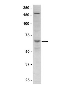
Western Blot Analysis: 2 μg/mL of this antibody detected AMPK α1 in RIPA lysates from non-stimulated A431 cells.

Immunohistochemistry (Paraffin) Analysis: 1:250 dilution of this antibody detected AMPK α1 in human gallbladder tissue sections.
Research Category
Signaling
Research Sub Category
Insulin/Energy Signaling

Quality

Evaluated by immunoblot in RIPA lysates from non-stimulated A431 cells.

Western Blot Analysis:
0.5-2 μg/mL of this antibody detected AMPK α1 in RIPA lysates from non-stimulated A431 cells.

Target description

~63 kDa observed; 64.00 kDa calculated. Uncharacterized bands may be observed in some lysate(s).

Linkage

Replaces: 04-323

Physical form

Format: Purified
Protein A chromatography
Protein A purified rabbit in buffer containing 0.1M Tris-Glycine, 0.15M NaCl, 0.05% Sodium Azide, pH 7.4.

Storage and Stability

Stable for 1 year at 2-8°C from date of receipt.

Analysis Note

Control
Positive Antigen Control: Catalog #12-301, non-stimulated A431 cell lysate. Add 2.5µL of 2-mercaptoethanol/100µL of lysate and boil for 5 minutes to reduce the preparation. Load 20µg of reduced lysate per lane for minigels.

Other Notes

Concentration: Please refer to the Certificate of Analysis for the lot-specific concentration.

Legal Information

UPSTATE is a registered trademark of Merck KGaA, Darmstadt, Germany

Disclaimer

Unless otherwise stated in our catalog or other company documentation accompanying the product(s), our products are intended for research use only and are not to be used for any other purpose, which includes but is not limited to, unauthorized commercial uses, in vitro diagnostic uses, ex vivo or in vivo therapeutic uses or any type of consumption or application to humans or animals.

Not finding the right product?  

Try our Product Selector Tool.

Storage Class Code

10 - Combustible liquids

WGK

WGK 1


Certificates of Analysis (COA)

Search for Certificates of Analysis (COA) by entering the products Lot/Batch Number. Lot and Batch Numbers can be found on a product’s label following the words ‘Lot’ or ‘Batch’.

Already Own This Product?

Find documentation for the products that you have recently purchased in the Document Library.

Visit the Document Library

Activation of AMPK?2 is not crucial for mitochondrial uncoupling-induced metabolic effects but required to maintain skeletal muscle integrity.
Ost, M; Werner, F; Dokas, J; Klaus, S; Voigt, A
Testing null
Aging elevates basal adenosine monophosphate-activated protein kinase (AMPK) activity and eliminates hypoxic activation of AMPK in mouse liver.
Jacob D Mulligan, Asensio A Gonzalez, Reetu Kumar, Ashley J Davis, Kurt W Saupe
The Journals of Gerontology. Series A, Biological Sciences and Medical Sciences null
Increased estrogen receptor ? in adipose tissue is associated with increased intracellular and reduced circulating adiponectin protein levels in aged female rats.
Nanette J Tomicek,Timothy S Lancaster,Donna H Korzick
Gender Medicine null
Miki Tadaishi et al.
American journal of physiology. Endocrinology and metabolism, 300(2), E341-E349 (2010-11-26)
There are three isoforms of peroxisome proliferator-activated receptor-γ coactivator 1α (PGC-1α) mRNA, which promotes mitochondrial biogenesis in skeletal muscles. Compared with PGC-1α-a mRNA, PGC-1α-b or PGC-1α-c mRNA is transcribed by a different exon 1 of the PGC-1α gene. In this
Thermogenic activation induces FGF21 expression and release in brown adipose tissue.
Hondares E, Iglesias R, Giralt A, Gonzalez FJ, Giralt M, Mampel T, Villarroya F
The Journal of Biological Chemistry null

Our team of scientists has experience in all areas of research including Life Science, Material Science, Chemical Synthesis, Chromatography, Analytical and many others.

Contact Technical Service