Skip to Content
MilliporeSigma
All Photos(1)

Documents

281M-9

Sigma-Aldrich

MART-1 (Melan A) (M2-7C10) Mouse Monoclonal Antibody

clone M2-7C10, unconjugated, Cell Marque

Sign Into View Organizational & Contract Pricing


About This Item

UNSPSC Code:
12352203
NACRES:
NA.41

biological source

mouse

Quality Level

100
500

conjugate

unconjugated

antibody form

culture supernatant

antibody product type

primary antibodies

clone

M2-7C10, monoclonal

description

For In Vitro Diagnostic Use in Select Regions (See Chart)

form

buffered aqueous solution

species reactivity

human

packaging

vial of 0.1 mL concentrate (281M-94)
vial of 0.5 mL concentrate (281M-95)
bottle of 1.0 mL predilute (281M-97)
vial of 1.0 mL concentrate (281M-96)
bottle of 7.0 mL predilute (281M-98)

manufacturer/tradename

Cell Marque

technique(s)

immunohistochemistry (formalin-fixed, paraffin-embedded sections): 1:100-1:500

isotype

IgG2bκ

control

melanoma

shipped in

wet ice

storage temp.

2-8°C

visualization

cytoplasmic

Gene Information

human ... MLANA(2315)

General description

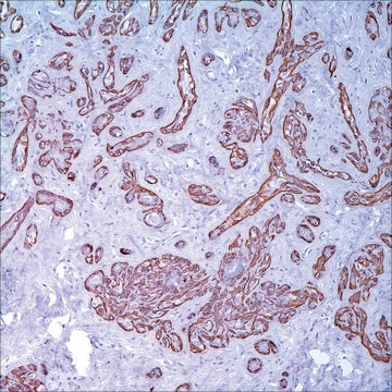
MART-1 (also known as Melan A) is a melanocyte differentiation antigen. It is present in melanocytes of normal skin, retina, nevi, and in more than 85% of melanomas. This antibody is very useful in establishing the diagnosis of metastatic melanomas.

Linkage

MART-1 Positive Control Slides, Product No. 281S, are available for immunohistochemistry (formalin-fixed, paraffin-embedded sections).

Physical form

Solution in Tris Buffer, pH 7.3-7.7, with 1% BSA and <0.1% Sodium Azide

Preparation Note

Download the IFU specific to your product lot and formatNote: This requires a keycode which can be found on your packaging or product label.

Other Notes

For Technical Service please contact: 800-665-7284 or email: service@cellmarque.com

Legal Information

Cell Marque is a trademark of Merck KGaA, Darmstadt, Germany

Not finding the right product?  

Try our Product Selector Tool.


Certificates of Analysis (COA)

Search for Certificates of Analysis (COA) by entering the products Lot/Batch Number. Lot and Batch Numbers can be found on a product’s label following the words ‘Lot’ or ‘Batch’.

Already Own This Product?

Find documentation for the products that you have recently purchased in the Document Library.

Visit the Document Library

R Bergman et al.
Journal of the American Academy of Dermatology, 42(3), 496-500 (2000-02-25)
The histopathologic differential diagnosis of Spitz nevus (SN) from malignant melanoma (MM) may be difficult. We attempted to elucidate the pattern of expression of a newly recognized melanocyte-specific melanosomal protein MART-1 in routinely processed specimens of SNs, MMs, and ordinary
Z Orosz
Histopathology, 34(6), 517-525 (1999-06-26)
The purpose of this study was to test different malignant non-melanocytic tumours with the commercially available antibody Melan-A to examine its diagnostic specificity and to compare the S100, Melan-A and HMB-45 reactivity in various melanocytic lesions. Seventy-three benign and malignant
T Kageshita et al.
Journal of immunotherapy (Hagerstown, Md. : 1997), 20(6), 460-465 (1997-12-31)
Twenty-eight primary and 29 metastatic melanoma lesions and 18 pigmented nevi lesions were analyzed by using the immunoperoxidase reaction with anti-MART-1 and anti-gp100 monoclonal antibodies (mAbs). The MART-1 was expressed in 28, 29, and 18, and gp100 was expressed in
P A Fetsch et al.
Cancer, 87(1), 37-42 (1999-03-30)
HMB-45, an antibody directed against a premelanosome glycoprotein, has thus far been considered the most specific antibody for the immunocytochemical substantiation of the diagnosis of malignant melanoma (MM). A recently described antigen, MART-1, is a transmembrane protein that is present
S Mocellin et al.
Journal of immunotherapy (Hagerstown, Md. : 1997), 24(6), 447-458 (2002-01-05)
Assessment of antigen expression by solid tumors has relied predominantly on immunohistochemistry, flow cytometry, and more recently quantitative real-time polymerase chain reaction. However, all these techniques present intrinsic limits. The laser scanning cytometer, by combining the properties of light and

Articles

IHC antibodies enhance dermatopathology beyond H&E stained slides, improving techniques and applications for dermatological research.

Related Content

Trusted IVDR-ready supplier of high-quality Cell Marque™ antibodies for diagnostic pathology and IHC, ensuring reliability and expert support.

Our team of scientists has experience in all areas of research including Life Science, Material Science, Chemical Synthesis, Chromatography, Analytical and many others.

Contact Technical Service