Skip to Content
MilliporeSigma
All Photos(2)

Documents

MAB5406B

Sigma-Aldrich

Anti-GAD67 Antibody, clone 1G10.2, Biotin Conjugate

clone 1G10.2, from mouse, biotin conjugate

Synonym(s):

Glutamate decarboxylase 1, 67 kDa glutamic acid decarboxylase, GAD-67, Glutamate decarboxylase 67 kDa isoform

Sign Into View Organizational & Contract Pricing


About This Item

UNSPSC Code:
12352203
eCl@ss:
32160702
NACRES:
NA.41

biological source

mouse

Quality Level

conjugate

biotin conjugate

antibody form

purified immunoglobulin

antibody product type

primary antibodies

clone

1G10.2, monoclonal

species reactivity

rat

species reactivity (predicted by homology)

human (based on 100% sequence homology), mouse (based on 100% sequence homology)

technique(s)

immunocytochemistry: suitable
immunohistochemistry: suitable

isotype

IgG2a

NCBI accession no.

UniProt accession no.

shipped in

wet ice

target post-translational modification

unmodified

Gene Information

human ... GAD1(2571)

General description

Gutamic acid decarboxylase (GAD; E.C. 4.1.1.15) is the enzyme responsible for the conversion of glutamic acid to gamma-aminobutyric acid (GABA), the major inhibitory transmitter in higher brain regions, and putative paracrine hormone in pancreatic islets. Two molecular forms of GAD (65 kDa and 67 kDa, 64% aa identity between forms) are highly conserved and both forms are expressed in the CNS, pancreatic islet cells, testis, oviduct and ovary. The isoforms are regionally distributed cytoplasmically in the brains of rats and mice (Sheikh, 1999). GAD65 is an amphiphilic, membrane-anchored protein (585 a.a.), encoded on human chromosome 10, and is responsible for vesicular GABA production. GAD67 is cytoplasmic (594 a.a.), encoded on chromosome 2, and seems to be responsible for significant cytoplasmic GABA production. GAD expression changes during neural development in rat spinal cord. GAD65 is expressed transiently in commissural axons around E13 but is down regulated the next day while GAD67 expression increases mostly in the somata of those neurons (Phelps, 1999). In mature rat pancreas, GAD65 and GAD67 appear to be differentially localized, GAD65 primarily in insulin-containing beta cells and GAD67 in glucagon-containing (A) cells (Li, 1995). GAD67 expression seems to be particularly plastic and can change in response to experimental manipulation (for example neuronal stimulation or transection) or disease progression and emergent disorders like schizophrenia (Volk, 2000). Colocalization of the two GAD isoforms also shows changes in GAD65/GAD67 distributions correlated with certain disease states such as IDDM and SMS.

Specificity

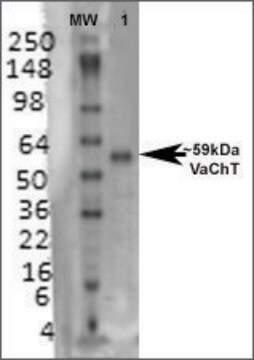
Reacts with GAD67. No detectable cross reactivity with GAD65 by Western blot on rat brain lysate.

Immunogen

Recombinant GAD67 protein

Application

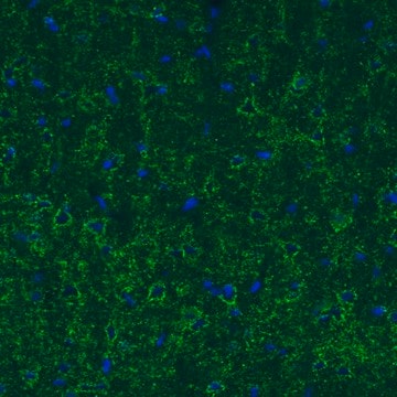
Immunocytochemistry Analysis: A 1:100 dilution from a representative lot detected GAD67 in rat E18 primary cortex cells.
Research Category
Neuroscience
Research Sub Category
Developmental Neuroscience
This Anti-GAD Antibody, clone 1G10.2, Biotin Conjugate is validated for use in IHC for the detection of GAD67. See below for more information about this anti GAD antibody.

Quality

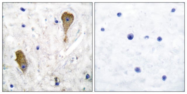
Evaluated by Immunohistochemistry in rat cortex tissue.

Immunohistochemistry Analysis: A 1:25 dilution of this antibody detected GAD67 in rat cortex tissue.

Target description

~67 kDa

Physical form

Protein A purified
Purified mouse monoclonal IgG2a conjugated to Biotin in PBS with 0.1% sodium azide and 15 mg/mL BSA.

Storage and Stability

Maintain refrigerated at 2-8 °C in undiluted aliquots for up to 6 months from date of receipt.

Analysis Note

Control
Rat cortex tissue

Disclaimer

Unless otherwise stated in our catalog or other company documentation accompanying the product(s), our products are intended for research use only and are not to be used for any other purpose, which includes but is not limited to, unauthorized commercial uses, in vitro diagnostic uses, ex vivo or in vivo therapeutic uses or any type of consumption or application to humans or animals.

Not finding the right product?  

Try our Product Selector Tool.

Storage Class

12 - Non Combustible Liquids

wgk_germany

WGK 2

flash_point_f

Not applicable

flash_point_c

Not applicable


Certificates of Analysis (COA)

Search for Certificates of Analysis (COA) by entering the products Lot/Batch Number. Lot and Batch Numbers can be found on a product’s label following the words ‘Lot’ or ‘Batch’.

Already Own This Product?

Find documentation for the products that you have recently purchased in the Document Library.

Visit the Document Library

Jose A Fernandez-Leon et al.
eLife, 10 (2021-12-17)
The recollection of environmental cues associated with threat or reward allows animals to select the most appropriate behavioral responses. Neurons in the prelimbic (PL) cortex respond to both threat- and reward-associated cues. However, it remains unknown whether PL regulates threat-avoidance
Jean-Christophe Delpech et al.
Science translational medicine, 13(611), eabe8455-eabe8455 (2021-09-16)
Abnormally phosphorylated tau, an early neuropathologic marker of Alzheimer’s disease (AD), first occurs in the brain’s entorhinal cortex layer II (ECII) and then spreads to the CA1 field of the hippocampus. Animal models of tau propagation aiming to recapitulate this
V Korzhova et al.
Communications biology, 4(1), 1368-1368 (2021-12-09)
Alzheimer's disease (AD) is associated with aberrant neuronal activity, which is believed to critically determine disease symptoms. How these activity alterations emerge, how stable they are over time, and whether cellular activity dynamics are affected by the amyloid plaque pathology
Hao-Yu Zou et al.
The Journal of clinical investigation, 132(8) (2022-04-16)
Mushroom spine loss and calcium dyshomeostasis are early hallmark events of age-related neurodegeneration, such as Alzheimer's disease (AD), that are connected with neuronal hyperactivity in early pathology of cognitive brain areas. However, it remains elusive how these key events are
Zhi Ruan et al.
Brain : a journal of neurology, 144(1), 288-309 (2020-11-28)
Extracellular vesicles are highly transmissible and play critical roles in the propagation of tau pathology, although the underlying mechanism remains elusive. Here, for the first time, we comprehensively characterized the physicochemical structure and pathogenic function of human brain-derived extracellular vesicles

Our team of scientists has experience in all areas of research including Life Science, Material Science, Chemical Synthesis, Chromatography, Analytical and many others.

Contact Technical Service