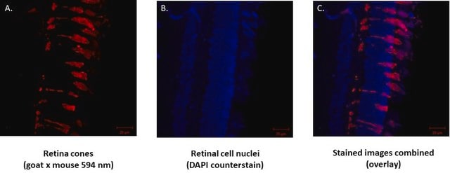
Detect Chx10 using this Anti-Chx10 Antibody, N-terminus validated for use in WB.
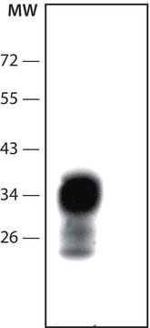
Immunoblotting: 0.5-1.0 μg/mL. Reacts with the 46 kD protein in rat and mouse retinal tissue lysate.
Optimal working dilutions must be determined by the end user.
Research Category Neuroscience
Research Sub Category Developmental Neuroscience
Neuronal & Glial Markers
Physical form
Format: Purified
Purified immunoglobulin. Liquid in PBS containing 0.08% sodium azide.
Storage and Stability
Maintain at -20°C in undiluted aliquots for up to 6 months after date of receipt. Avoid repeated freeze/thaw cycles.
Other Notes
Concentration: Please refer to the Certificate of Analysis for the lot-specific concentration.
Legal Information
CHEMICON is a registered trademark of Merck KGaA, Darmstadt, Germany
Disclaimer
Unless otherwise stated in our catalog or other company documentation accompanying the product(s), our products are intended for research use only and are not to be used for any other purpose, which includes but is not limited to, unauthorized commercial uses, in vitro diagnostic uses, ex vivo or in vivo therapeutic uses or any type of consumption or application to humans or animals.
Not finding the right product?
Try our Product Selector Tool.
Storage Class
10 - Combustible liquids
wgk_germany
WGK 2
flash_point_f
Not applicable
flash_point_c
Not applicable
Certificates of Analysis (COA)
Search for Certificates of Analysis (COA) by entering the products Lot/Batch Number. Lot and Batch Numbers can be found on a product’s label following the words ‘Lot’ or ‘Batch’.
Already Own This Product?
Find documentation for the products that you have recently purchased in the Document Library.
Effective derivation of three-dimensional (3D) retinal tissue from human-induced pluripotent stem cells (hiPSCs) could provide models for drug screening and facilitate patient-specific retinal cell replacement therapy. However, some hiPSC lines cannot undergo 3D self-organization and show inadequate differentiation efficiency to
Proceedings of the National Academy of Sciences of the United States of America, 116(22), 10824-10833 (2019-05-11)
Rod and cone photoreceptors are light-sensing cells in the human retina. Rods are dominant in the peripheral retina, whereas cones are enriched in the macula, which is responsible for central vision and visual acuity. Macular degenerations affect vision the most
Urine cells, a body trash, have been successfully reprogrammed into human induced pluripotent stem cells (U-hiPSCs) which hold a huge promise in regenerative medicine. However, it is unknown whether or to what extent U-hiPSCs can generate retinal cells so far.
We intend to identify marker genes with differential gene expression (DEG) and RGC subtypes in cultures of human-induced pluripotent stem cell (iPSC)-derived retinal ganglion cells. Single-cell sequencing was performed on mature and functional iPSC-RGCs at day 40 using Chromium Single
The telencephalon and eye in mammals are originated from adjacent fields at the anterior neural plate. Morphogenesis of these fields generates telencephalon, optic-stalk, optic-disc, and neuroretina along a spatial axis. How these telencephalic and ocular tissues are specified coordinately to
Our team of scientists has experience in all areas of research including Life Science, Material Science, Chemical Synthesis, Chromatography, Analytical and many others.