Przejdź do zawartości
Merck
HomeCancer ResearchPrestige Antibodies® in Breast Cancer Research

Prestige Antibodies® in Breast Cancer Research

prestige antibodies-logo
prestige antibodies

The Human Protein Atlas

proteinatlas.org

The Human Protein Atlas is Characterizing the Human Proteome

The Human Protein Atlas (HPA) project was initiated in 2003 by Swedish researchers, headed by Professor Mathias Uhlén, and funded by the Knut and Alice Wallenberg Foundation.1,2 It is a unique world leading effort performing systematic exploration of the human proteome using antibodies.

The aim of the HPA project is to present an expression map of the complete human proteome. To accomplish this, highly specific polyclonal antibodies are developed to all protein coding human genes and protein profiling is established in a multitude of tissues and cells using tissue arrays. Applications applied are immunohistochemistry (IHC), Western blot (WB) analysis, protein array assay and immunofluorescent based confocal microscopy (ICC-IF).

The Human Protein Atlas, November 2014

The 13th version of the Human Protein Atlas, released in November 2014, presents a tissue-based map of the complete human proteome. The extensive amount of data is divided into four separate 'sub atlases': the Tissue Atlas, the Cancer Atlas, the Subcell Atlas and the Cell Line Atlas. For all proteins represented in the Tissue Atlas, the expression profiles are based on IHC analysis on a large number of human tissues. The presentation of protein expression data in correlation to RNA sequencing data for each gene has now been included. In the Cancer Atlas, differentially expressed genes in several cancers can be studied, while the Subcell Atlas presents subcellular localization by confocal microscopy. Additional information about protein expression in common cell lines is included in the Cell Line Atlas, which has become an appreciated toolbox for research.

Tissue microarrays containing samples from 48 different normal human tissues, 20 different cancer types and 44 different human cell lines are utilized within the project. The 48 normal tissues are present in triplicate samples and represent 82 different cell types. All normal tissue images have undergone pathology-based annotation of expression levels and are displayed on the normal Tissue Atlas presenting information regarding the expression profiles of human genes both on mRNA and protein level. The mRNA expression data is derived from deep sequencing of RNA (RNA-Seq) from 27 major different normal tissue types.

The Cancer Atlas contains gene expression data based on protein expression patterns in a multitude of human cancer specimens. Altogether 216 different cancer samples, corresponding to the 20 most common forms of human cancer, have been analyzed for all included genes. All cancer tissue images have been manually annotated by pathologists and just as for the normal Tissue Atlas, protein data includes protein expression levels corresponding to 16.621 genes for which there are available antibodies.

Validation in Breast Tissue Samples and Cell Lines

IHC images from normal breast samples from three different individuals are available for each antibody in the normal Tissue Atlas. In addition, for each antibody, breast tumor samples from up to 12 patients in duplicates are presented in the Cancer Atlas and for the majority of the antibodies, also images from the MCF-7 and SK-BR-3 breast cell lines in the Cell Line Atlas.

References

1.
Uhlen M, Oksvold P, Fagerberg L, Lundberg E, Jonasson K, Forsberg M, Zwahlen M, Kampf C, Wester K, Hober S, et al. 2010. Towards a knowledge-based Human Protein Atlas. Nat Biotechnol. 28(12):1248-1250. https://doi.org/10.1038/nbt1210-1248
2.
Uhlen M, Ponten F, Lindskog C. 2015. Charting the human proteome: Understanding disease using a tissue-based atlas. Science. 347(6227):1274-1274. https://doi.org/10.1126/science.347.6227.1274-c

Prestige Antibodies® Powered by Atlas Antibodies

Prestige Antibodies – the Building Blocks of HPA

The uniqueness and low cross reactivity of Prestige Polyclonals to other proteins are due to a thorough selection of antigen regions, affinity purification on the recombinant antigen, validation using several methods and a stringent approval process.

Development

The Prestige Antibodies are developed against recombinant human Protein Epitope Signature Tags (PrESTs) of approximately 50 to 150 amino acids. These protein fragments are designed, using a proprietary software, to contain unique epitopes present in the native protein suitable for triggering the generation of antibodies of high specificity. This is achieved by a complete human genome scanning to ensure that PrESTs with the lowest homology to other human proteins are used as antigens.

Approval

The approval of the Prestige Antibodies relies on a combined validation of the experimental results using IHC, WB or ICC-IF, from RNA sequencing and from information obtained via bioinformatics prediction methods and literature. Since the literature is often inconclusive, an important objective of the HPA project has been to generate paired antibodies with non-overlapping epitopes towards the same protein target, allowing the results and validation of one antibody to be used to validate the other one.

Prestige Antibodies Catalog

Today, there are more than 17,000 Prestige Polyclonals with 2,000 new antibodies added each year.

The antibodies developed and characterized within the Human Protein Atlas project are available to the scientific community  as Prestige Antibodies and Atlas Antibodies.


Monoclonal Antibody Development

Prestige Antibodies also include a selected number of mouse monoclonal antibodies. The monoclonal catalog is regularly expanding with new products every year.

Unique Features

Special care is taken in offering clones recognizing only unique nonoverlapping epitopes and/or isotypes. Using the same stringent PrEST production process and characterization procedure as for the polyclonals, the monoclonal antibodies offer outstanding performance in approved applications, together with defined specificity, secured continuity and stable supply. In general they also permit high working dilutions and contribute to more standardized assay procedures.

Clone Selection

Functional characterization is performed on a large number of ELISA positive cell supernatants to select the optimal clones for each application prior to subcloning and expansion of selected hybridomas.

Epitope Mapping

Clones are epitope-mapped using synthetic overlapping peptides in a bead-based array format for selection of clones with nonoverlapping epitopes only.

Isotyping

All monoclonal antibodies are isotyped to allow for multiplexing using isotype-specific secondary antibodies.

Hybridoma Cell Cultivation

Atlas Antibodies use in vitro methods for the production scale-up phase thus replacing the use of mice for production of ascites fluid.

Antibody Characterization

The characterization of Prestige Monoclonal Antibodies starts with an extensive literature search to select the most relevant and clinically significant tissues to use for IHC characterization. Often there are more than one tissue type displayed in the IHC application data for each antibody. In addition to positive stained tissue, a negative control tissue staining is also displayed and if relevant, clinical cancer tissue staining.

The Western blot (WB) characterization includes results from endogenous human cell or tissue protein lysates or optionally recombinant full-length human protein lysates.

Each monoclonal antibody is thus supplied with the most relevant characterization data for its specific target.

The product numbers of all Prestige Polyclonals start with ”HPA” and of monoclonal antibodies with “AMAB”.


Clinical Markers (ESR1, HER2, Ki67, PGR)

Established Clinical Breast Cancer Markers

1.
Pereira CBL, Leal MF, de Souza CRT, Montenegro RC, Rey JA, Carvalho AA, Assumpção PP, Khayat AS, Pinto GR, Demachki S, et al. Prognostic and Predictive Significance of MYC and KRAS Alterations in Breast Cancer from Women Treated with Neoadjuvant Chemotherapy. PLoS ONE. 8(3):e60576. https://doi.org/10.1371/journal.pone.0060576
2.
Camilleri M, Carlson P, Zinsmeister AR, McKinzie S, Busciglio I, Burton D, Zucchelli M, D'Amato M. 2010. Neuropeptide S Receptor Induces Neuropeptide Expression and Associates With Intermediate Phenotypes of Functional Gastrointestinal Disorders. Gastroenterology. 138(1):98-107.e4. https://doi.org/10.1053/j.gastro.2009.08.051
3.
Roca H, Craig MJ, Ying C, Varsos ZS, Czarnieski P, Alva AS, Hernandez J, Fuller D, Daignault S, Healy PN, et al. 2011. IL-4 induces proliferation in prostate cancer PC3?cells under nutrient-depletion stress through the activation of the JNK-pathway and survivin upregulation. J. Cell. Biochem..n/a-n/a. https://doi.org/10.1002/jcb.24025
HER2/ERBB2

Immunohistochemical staining of human breast tumour using Anti-HER2 (AMAb90627) shows strong membranous (combined with moderate cytoplasmic) positivity in tumour cells in HER2-positive ductal carcinoma, while HER2-negative ductal carcinoma shows no membranous positivity. By Western Blot analysis, HER2 is detected in the breast cancer cell line SK-BR-3.

Progesteron Receptor

Progesteron Receptor

IHC staining using the Anti-PGR antibody (HPA004751) in normal human corpus (uterine) tissue shows strong nuclear positivity in glandular cells. In the presented breast cancer sample, the staining of tumor cells is also nuclear. ICC-IF shows nuclear staining in U-251MG cells.

Estrogen Receptor

Estrogen Receptor

The Anti-ESR1 antibody (HPA000449) shows distinct nuclear positivity in glandular cells in human breast tissue and in tumor cells in breast cancer samples using IHC.

Estrogen Receptor-2

IHC staining using the Anti-ESR1 antibody (HPA000450) shows strong nuclear positivity in glandular and stromal cells of human corpus, uterine tissue and in tumor cells in breast cancer.

Ki67

Ki67

The Anti-MKI67 antibody (HPA000451) shows strong nuclear positivity in a fraction of cells in the reaction center in human lymph node using IHC. In breast cancer, the staining of tumor cells is also nuclear and by ICC-IF, staining of the human cell line U-2OS shows positivity in nucleoli.

ki67-2

IHC staining of human tonsil tissue using the Anti-MKI67 antibody (HPA001164) shows nuclear staining of reaction center cells. In tumor cells in breast cancer, the staining is mainly nuclear and in U-2OS cells, using ICC-IF, nucleoli show strong positivity.

ki67-3

IHC staining of lymph node in human colon shows strong nuclear and nucleolar immunoreactivity in the reaction centrum cells using the monoclonal Anti-MKI67 antibody (AMAb90870). In uterus, nuclear positivity in a subset of glandular cells is shown.


Antibodies used in Breast Cancer Research

In this section, antibodies are selected either on a reference/article-basis or on breast cancer relevance for the corresponding target protein.

BRCA1

brca1-1

The Anti-BRCA1 antibody (HPA034966) shows positivity in glandular cells in normal human breast tissue and in tumor cells in breast cancer samples using IHC.

brca1-2

IHC staining using the Anti-BRCA2 antibody (HPA026815) in normal human breast tissue shows positivity in glandular cells. In breast cancer, nuclear staining of tumor cells is shown.

ACAT1

acat1

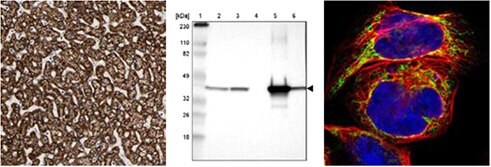
Immunohistochemical staining of human liver tissue using Anti-ACAT1 (HPA004428) shows strong cytoplasmic positivity in hepatocytes. By Western blot analysis, ACAT1 is detected in the human cell lines RT-4 and U251-MG and in liver and tonsil tissue lysates. By ICC-IF in the human cell line A-431, positivity is shown in mitochondria.

CD44

cd44

Immunohistochemical staining of human esophagus tissue using Anti-CD44 (HPA005785) shows strong strong cytoplasmic and membranous positivity in squamous epithelial cells. By Western Blot analysis, CD44 is detected in the human cell line U-251MG. ICC-IF in the human cell line U-251MG shows positivity in plasma membrane.

* WB both in human and rodent samples

References

1.
Sanchez-Alvarez R, Martinez-Outschoorn UE, Lin Z, Lamb R, Hulit J, Howell A, Sotgia F, Rubin E, Lisanti MP. 2013. Ethanol exposure induces the cancer-associated fibroblast phenotype and lethal tumor metabolism. Cell Cycle. 12(2):289-301. https://doi.org/10.4161/cc.23109
2.
Martinez-Outschoorn UE, Lin Z, Whitaker-Menezes D, Howell A, Lisanti MP, Sotgia F. 2012. Ketone bodies and two-compartment tumor metabolism: Stromal ketone production fuels mitochondrial biogenesis in epithelial cancer cells. Cell Cycle. 11(21):3956-3963. https://doi.org/10.4161/cc.22136
3.
Martinez-Outschoorn UE, Lin Z, Whitaker-Menezes D, Howell A, Sotgia F, Lisanti MP. 2012. Ketone body utilization drives tumor growth and metastasis. Cell Cycle. 11(21):3964-3971. https://doi.org/10.4161/cc.22137
4.
Chang HT, Olson L, Schwartz KA. 2013. Ketolytic and glycolytic enzymatic expression profiles in malignant gliomas: implication for ketogenic diet therapy. Nutrition & Metabolism. 10(1):47. https://doi.org/10.1186/1743-7075-10-47
5.
Hrstka R, Brychtova V, Fabian P, Vojtesek B, Svoboda M. 2013. AGR2 Predicts Tamoxifen Resistance in Postmenopausal Breast Cancer Patients. Disease Markers. 35207-212. https://doi.org/10.1155/2013/761537
6.
O´Leary PC, Penny SA, Dolan RT, Kelly CM, Madden SF, Rexhepaj E, Brennan DJ, McCann AH, Pontén F, Uhlén M, et al. 2013. Systematic antibody generation and validation via tissue microarray technology leading to identification of a novel protein prognostic panel in breast cancer. BMC Cancer. 13(1): https://doi.org/10.1186/1471-2407-13-175
7.
de Boniface J, Mao Y, Schmidt-Mende J, Kiessling R, Poschke I. 2012. Expression patterns of the immunomodulatory enzyme arginase 1 in blood, lymph nodes and tumor tissue of early-stage breast cancer patients. OncoImmunology. 1(8):1305-1312. https://doi.org/10.4161/onci.21678
8.
Lucki NC, Li D, Bandyopadhyay S, Wang E, Merrill AH, Sewer MB. 2012. Acid Ceramidase (ASAH1) Represses Steroidogenic Factor 1-Dependent Gene Transcription in H295R Human Adrenocortical Cells by Binding to the Receptor. Molecular and Cellular Biology. 32(21):4419-4431. https://doi.org/10.1128/mcb.00378-12
9.
Liang Y, Wu H, Lei R, Chong RA, Wei Y, Lu X, Tagkopoulos I, Kung S, Yang Q, Hu G, et al. 2012. Transcriptional Network Analysis Identifies BACH1 as a Master Regulator of Breast Cancer Bone Metastasis. J. Biol. Chem.. 287(40):33533-33544. https://doi.org/10.1074/jbc.m112.392332
10.
Scheper M, Almubarak H, Jones A, Chaisuparat R, Zhang M, Meiller T. 2011. Zoledronic acid directly suppresses cell proliferation and induces apoptosis in highly tumorigenic prostate and breast cancers. J Carcinog. 10(1):2. https://doi.org/10.4103/1477-3163.75723
11.
Brunquell C, Biliran H, Jennings S, Ireland SK, Chen R, Ruoslahti E. 2012. TLE1 Is an Anoikis Regulator and Is Downregulated by Bit1 in Breast Cancer Cells. Molecular Cancer Research. 10(11):1482-1495. https://doi.org/10.1158/1541-7786.mcr-12-0144
12.
Karmali PP, Brunquell C, Tram H, Ireland SK, Ruoslahti E, Biliran H. Metastasis of Tumor Cells Is Enhanced by Downregulation of Bit1. PLoS ONE. 6(8):e23840. https://doi.org/10.1371/journal.pone.0023840
13.
Vermeulen JF, van Brussel AS, van der Groep P, Morsink FH, Bult P, van der Wall E, van Diest PJ. 2012. Immunophenotyping invasive breast cancer: paving the road for molecular imaging. BMC Cancer. 12(1): https://doi.org/10.1186/1471-2407-12-240
14.
Davidson B, Stavnes HT, Holth A, Chen X, Yang Y, Shih I, Wang T. 2011. Gene expression signatures differentiate ovarian/peritoneal serous carcinoma from breast carcinoma in effusions. 15(3):535-544. https://doi.org/10.1111/j.1582-4934.2010.01019.x
15.
Vermeulen JF, Kornegoor R, van der Wall E, van der Groep P, van Diest PJ. Differential Expression of Growth Factor Receptors and Membrane-Bound Tumor Markers for Imaging in Male and Female Breast Cancer. PLoS ONE. 8(1):e53353. https://doi.org/10.1371/journal.pone.0053353
16.
Tafreshi NK, Bui MM, Bishop K, Lloyd MC, Enkemann SA, Lopez AS, Abrahams D, Carter BW, Vagner J, Grobmyer SR, et al. 2012. Noninvasive Detection of Breast Cancer Lymph Node Metastasis Using Carbonic Anhydrases IX and XII Targeted Imaging Probes. Clinical Cancer Research. 18(1):207-219. https://doi.org/10.1158/1078-0432.ccr-11-0238
17.
Vazquez-Martin A, Oliveras-Ferraros C, Cufí S, Del Barco S, Martin-Castillo B, Menendez JA. 2010. Metformin regulates breast cancer stem cello ntogeny by transcriptional regulation of the epithelial-mesenchymal transition (EMT) status. Cell Cycle. 9(18):3831-3838. https://doi.org/10.4161/cc.9.18.13131
18.
Baccelli I, Schneeweiss A, Riethdorf S, Stenzinger A, Schillert A, Vogel V, Klein C, Saini M, Bäuerle T, Wallwiener M, et al. 2013. Identification of a population of blood circulating tumor cells from breast cancer patients that initiates metastasis in a xenograft assay. Nat Biotechnol. 31(6):539-544. https://doi.org/10.1038/nbt.2576
19.
Petit V, Massonnet G, Maciorowski Z, Touhami J, Thuleau A, Némati F, Laval J, Château-Joubert S, Servely J, Vallerand D, et al. 2013. Optimization of tumor xenograft dissociation for the profiling of cell surface markers and nutrient transporters. Lab Invest. 93(5):611-621. https://doi.org/10.1038/labinvest.2013.44
20.
Twarock S, Röck K, Sarbia M, Weber A, Jänicke R, Fischer J. Synthesis of hyaluronan in oesophageal cancer cells is uncoupled from the prostaglandin-cAMP pathway. 157(2):234-243. https://doi.org/10.1111/j.1476-5381.2009.00138.x
21.
Asplund A, Gry Björklund M, Sundquist C, Strömberg S, Edlund K, Östman A, Nilsson P, Pontén F, Lundeberg J. Expression profiling of microdissected cell populations selected from basal cells in normal epidermis and basal cell carcinoma. 158(3):527-538. https://doi.org/10.1111/j.1365-2133.2007.08418.x
22.
Teleki I, Krenacs T, Szasz MA, Kulka J, Wichmann B, Leo C, Papassotiropoulos B, Riemenschnitter C, Moch H, Varga Z. 2013. The potential prognostic value of connexin 26 and 46 expression in neoadjuvant-treated breast cancer. BMC Cancer. 13(1): https://doi.org/10.1186/1471-2407-13-50
23.
Kiflemariam S, Andersson S, Asplund A, Pontén F, Sjöblom T. Scalable In Situ Hybridization on Tissue Arrays for Validation of Novel Cancer and Tissue-Specific Biomarkers. PLoS ONE. 7(3):e32927. https://doi.org/10.1371/journal.pone.0032927
24.
Nodin B, Fridberg M, Uhlén M, Jirström K. 2012. Discovery of dachshund 2 protein as a novel biomarker of poor prognosis in epithelial ovarian cancer. Journal of Ovarian Research. 5(1):6. https://doi.org/10.1186/1757-2215-5-6
25.
Sircoulomb F, Nicolas N, Ferrari A, Finetti P, Bekhouche I, Rousselet E, Lonigro A, Adélaïde J, Baudelet E, Esteyriès S, et al. 2011. ZNF703 gene amplification at 8p12 specifies luminal B breast cancer. EMBO Mol Med. 3(3):153-166. https://doi.org/10.1002/emmm.201100121
26.
Cawthorn TR, Moreno JC, Dharsee M, Tran-Thanh D, Ackloo S, Zhu PH, Sardana G, Chen J, Kupchak P, Jacks LM, et al. Proteomic Analyses Reveal High Expression of Decorin and Endoplasmin (HSP90B1) Are Associated with Breast Cancer Metastasis and Decreased Survival. PLoS ONE. 7(2):e30992. https://doi.org/10.1371/journal.pone.0030992
27.
Henke A, Grace OC, Ashley GR, Stewart GD, Riddick ACP, Yeun H, O?Donnell M, Anderson RA, Thomson AA. Stromal Expression of Decorin, Semaphorin6D, SPARC, Sprouty1 and Tsukushi in Developing Prostate and Decreased Levels of Decorin in Prostate Cancer. PLoS ONE. 7(8):e42516. https://doi.org/10.1371/journal.pone.0042516
28.
Hudson EP, Uhlen M, Rockberg J. 2012. Multiplex epitope mapping using bacterial surface display reveals both linear and conformational epitopes. Sci Rep. 2(1): https://doi.org/10.1038/srep00706
29.
Arabi A, Ullah K, Branca RM, Johansson J, Bandarra D, Haneklaus M, Fu J, Ariës I, Nilsson P, Den Boer ML, et al. 2012. Proteomic screen reveals Fbw7 as a modulator of the NF-?B pathway. Nat Commun. 3(1): https://doi.org/10.1038/ncomms1975
30.
Ito A, Mimae T, Yamamoto Y, Hagiyama M, Nakanishi J, Ito M, Hosokawa Y, Okada M, Murakami Y, Kondo T. 2012. Novel application for pseudopodia proteomics using excimer laser ablation and two-dimensional difference gel electrophoresis. Lab Invest. 92(9):1374-1385. https://doi.org/10.1038/labinvest.2012.98
31.
Holland DG, Burleigh A, Git A, Goldgraben MA, Perez?Mancera PA, Chin S, Hurtado A, Bruna A, Ali HR, Greenwood W, et al. 2011. ZNF703 is a common Luminal B breast cancer oncogene that differentially regulates luminal and basal progenitors in human mammary epithelium. EMBO Mol Med. 3(3):167-180. https://doi.org/10.1002/emmm.201100122
32.
Mulder J, Björling E, Jonasson K, Wernérus H, Hober S, Hökfelt T, Uhlén M. 2009. Tissue Profiling of the Mammalian Central Nervous System Using Human Antibody-based Proteomics. Mol Cell Proteomics. 8(7):1612-1622. https://doi.org/10.1074/mcp.m800539-mcp200
33.
Su D, Fu X, Fan S, Wu X, Wang X, Fu L, Dong X, Ni JJ, Fu L, Zhu Z, et al. 2012. Role of ERRF, a Novel ER-Related Nuclear Factor, in the Growth Control of ER-Positive Human Breast Cancer Cells. The American Journal of Pathology. 180(3):1189-1201. https://doi.org/10.1016/j.ajpath.2011.11.025
34.
Shubbar E, Helou K, Kovács A, Nemes S, Hajizadeh S, Enerbäck C, Einbeigi Z. 2013. High levels of ?-glutamyl hydrolase (GGH) are associated with poor prognosis and unfavorable clinical outcomes in invasive breast cancer. BMC Cancer. 13(1): https://doi.org/10.1186/1471-2407-13-47
35.
Liao YC, Ruan JW, Lua I, Li MH, Chen WL, Wang JRY, Kao RH, Chen JH. 2012. Overexpressed hPTTG1 promotes breast cancer cell invasion and metastasis by regulating GEF-H1/RhoA signalling. Oncogene. 31(25):3086-3097. https://doi.org/10.1038/onc.2011.476
36.
Cheng IK, Tsang BC, Lai KP, Ching AK, Chan AW, To K, Lai PB, Wong N. 2012. GEF-H1 over-expression in hepatocellular carcinoma promotes cell motility via activation of RhoA signalling. J. Pathol.. 228(4):575-585. https://doi.org/10.1002/path.4084
37.
Zibert JR, Wallbrecht K, Schön M, Mir LM, Jacobsen GK, Trochon-Joseph V, Bouquet C, Villadsen LS, Cadossi R, Skov L, et al. 2011. Halting angiogenesis by non-viral somatic gene therapy alleviates psoriasis and murine psoriasiform skin lesions. J. Clin. Invest.. 121(1):410-421. https://doi.org/10.1172/jci41295
38.
Smyth LG, O'Hurley G, O'Grady A, Fitzpatrick JM, Kay E, Watson RWG. 2010. Carbonic anhydrase IX expression in prostate cancer. Prostate Cancer Prostatic Dis. 13(2):178-181. https://doi.org/10.1038/pcan.2009.58
39.
Paatero I, Jokilammi A, Heikkinen PT, Iljin K, Kallioniemi O, Jones FE, Jaakkola PM, Elenius K. 2012. Interaction with ErbB4 Promotes Hypoxia-inducible Factor-1? Signaling. J. Biol. Chem.. 287(13):9659-9671. https://doi.org/10.1074/jbc.m111.299537
40.
Zbytek B, Peacock DL, Seagroves TN, Slominski A. 2013. Putative role of HIF transcriptional activity in melanocytes and melanoma biology. Dermato-Endocrinology. 5(2):239-251. https://doi.org/10.4161/derm.22678
41.
Hu Z, Huang G, Sadanandam A, Gu S, Lenburg ME, Pai M, Bayani N, Blakely EA, Gray JW, Mao J. 2010. The expression level of HJURP has an independent prognostic impact and predicts the sensitivity to radiotherapy in breast cancer. Breast Cancer Res. 12(2): https://doi.org/10.1186/bcr2487
42.
Shuaib M, Ouararhni K, Dimitrov S, Hamiche A. 2010. HJURP binds CENP-A via a highly conserved N-terminal domain and mediates its deposition at centromeres. Proceedings of the National Academy of Sciences. 107(4):1349-1354. https://doi.org/10.1073/pnas.0913709107
43.
de Tayrac M, Saikali S, Aubry M, Bellaud P, Boniface R, Quillien V, Mosser J. Prognostic Significance of EDN/RB, HJURP, p60/CAF-1 and PDLI4, Four New Markers in High-Grade Gliomas. PLoS ONE. 8(9):e73332. https://doi.org/10.1371/journal.pone.0073332
44.
Bjarnadottir O, Romero Q, Bendahl P, Jirström K, Rydén L, Loman N, Uhlén M, Johannesson H, Rose C, Grabau D, et al. 2013. Targeting HMG-CoA reductase with statins in a window-of-opportunity breast cancer trial. Breast Cancer Res Treat. 138(2):499-508. https://doi.org/10.1007/s10549-013-2473-6
45.
Jaraj SJ, Augsten M, Häggarth L, Wester K, Pontén F, Östman A, Egevad L. 2011. GAD1 is a biomarker for benign and malignant prostatic tissue. Scandinavian Journal of Urology and Nephrology. 45(1):39-45. https://doi.org/10.3109/00365599.2010.521189
46.
Liu H, Zhang W, Jia Y, Yu Q, Grau GE, Peng L, Ran Y, Yang Z, Deng H, Lou J. 2013. Single-cell clones of liver cancer stem cells have the potential of differentiating into different types of tumor cells. Cell Death Dis. 4(10):e857-e857. https://doi.org/10.1038/cddis.2013.340
47.
Guo L, Chen C, Shi M, Wang F, Chen X, Diao D, Hu M, Yu M, Qian L, Guo N. 2013. Stat3-coordinated Lin-28?let-7?HMGA2 and miR-200?ZEB1 circuits initiate and maintain oncostatin M-driven epithelial?mesenchymal transition. Oncogene. 32(45):5272-5282. https://doi.org/10.1038/onc.2012.573
48.
Possemato R, Marks KM, Shaul YD, Pacold ME, Kim D, Birsoy K, Sethumadhavan S, Woo H, Jang HG, Jha AK, et al. 2011. Functional genomics reveal that the serine synthesis pathway is essential in breast cancer. Nature. 476(7360):346-350. https://doi.org/10.1038/nature10350
49.
Maddocks ODK, Berkers CR, Mason SM, Zheng L, Blyth K, Gottlieb E, Vousden KH. 2013. Serine starvation induces stress and p53-dependent metabolic remodelling in cancer cells. Nature. 493(7433):542-546. https://doi.org/10.1038/nature11743
50.
Nilsson LM, Plym Forshell TZ, Rimpi S, Kreutzer C, Pretsch W, Bornkamm GW, Nilsson JA. Mouse Genetics Suggests Cell-Context Dependency for Myc-Regulated Metabolic Enzymes during Tumorigenesis. PLoS Genet. 8(3):e1002573. https://doi.org/10.1371/journal.pgen.1002573
51.
Salem AF, Whitaker-Menezes D, Howell A, Sotgia F, Lisanti MP. 2012. Mitochondrial biogenesis in epithelial cancer cells promotes breast cancer tumor growth and confers autophagy resistance. Cell Cycle. 11(22):4174-4180. https://doi.org/10.4161/cc.22376
52.
Wagoner MP, Gunsalus KTW, Schoenike B, Richardson AL, Friedl A, Roopra A. The Transcription Factor REST Is Lost in Aggressive Breast Cancer. PLoS Genet. 6(6):e1000979. https://doi.org/10.1371/journal.pgen.1000979
53.
Prada I, Marchaland J, Podini P, Magrassi L, D'Alessandro R, Bezzi P, Meldolesi J. 2011. REST/NRSF governs the expression of dense-core vesicle gliosecretion in astrocytes. 193(3):537-549. https://doi.org/10.1083/jcb.201010126
54.
Jögi A, Brennan DJ, Rydén L, Magnusson K, Fernö M, Stål O, Borgquist S, Uhlen M, Landberg G, Påhlman S, et al. 2009. Nuclear expression of the RNA-binding protein RBM3 is associated with an improved clinical outcome in breast cancer. Mod Pathol. 22(12):1564-1574. https://doi.org/10.1038/modpathol.2009.124
55.
Hjelm B, Brennan DJ, Zendehrokh N, Eberhard J, Nodin B, Gaber A, Pontén F, Johannesson H, Smaragdi K, Frantz C, et al. 2011. High nuclear RBM3 expression is associated with an improved prognosis in colorectal cancer. Prot. Clin. Appl.. 5(11-12):624-635. https://doi.org/10.1002/prca.201100020
56.
Ehlén Å, Brennan DJ, Nodin B, O'Connor DP, Eberhard J, Alvarado-Kristensson M, Jeffrey IB, Manjer J, Brändstedt J, Uhlén M, et al. 2010. Expression of the RNA-binding protein RBM3 is associated with a favourable prognosis and cisplatin sensitivity in epithelial ovarian cancer. Journal of Translational Medicine. 8(1):78. https://doi.org/10.1186/1479-5876-8-78
57.
Jonsson L, Gaber A, Ulmert D, Uhlén M, Bjartell A, Jirström K. 2011. High RBM3 expression in prostate cancer independently predicts a reduced risk of biochemical recurrence and disease progression. Diagn Pathol. 6(1): https://doi.org/10.1186/1746-1596-6-91
58.
Nodin B, Fridberg M, Jonsson L, Bergman J, Uhlén M, Jirström K. 2012. High MCM3 expression is an independent biomarker of poor prognosis and correlates with reduced RBM3 expression in a prospective cohort of malignant melanoma. Diagnostic Pathology. 7(1):82. https://doi.org/10.1186/1746-1596-7-82
59.
Jonsson L, Bergman J, Nodin B, Manjer J, Pontén F, Uhlén M, Jirström K. 2011. Low RBM3 protein expression correlates with tumour progression and poor prognosis in malignant melanoma: An analysis of 215 cases from the Malmö Diet and Cancer Study. Journal of Translational Medicine. 9(1):114. https://doi.org/10.1186/1479-5876-9-114
60.
Ehlén Õ, Nodin B, Rexhepaj E, Brändstedt J, Uhlén M, Alvarado-Kristensson M, Pontén F, Brennan DJ, Jirström K. 2011. RBM3-Regulated Genes Promote DNA Integrity and Affect Clinical Outcome in Epithelial Ovarian Cancer. Translational Oncology. 4(4):212-IN1. https://doi.org/10.1593/tlo.11106
61.
Hjelm B, Brennan DJ, Zendehrokh N, Eberhard J, Nodin B, Gaber A, Pontén F, Johannesson H, Smaragdi K, Frantz C, et al. 2011. High nuclear RBM3 expression is associated with an improved prognosis in colorectal cancer. Prot. Clin. Appl.. 5(11-12):624-635. https://doi.org/10.1002/prca.201100020
62.
Boman K, Segersten U, Ahlgren G, Eberhard J, Uhlén M, Jirström K, Malmström P. 2013. Decreased expression of RNA-binding motif protein 3 correlates with tumour progression and poor prognosis in urothelial bladder cancer. BMC Urol. 13(1): https://doi.org/10.1186/1471-2490-13-17
63.
Nodin B, Fridberg M, Jonsson L, Bergman J, Uhlén M, Jirström K. 2012. High MCM3 expression is an independent biomarker of poor prognosis and correlates with reduced RBM3 expression in a prospective cohort of malignant melanoma. Diagnostic Pathology. 7(1):82. https://doi.org/10.1186/1746-1596-7-82
64.
Telikicherla D, Marimuthu A, Kashyap M, Ramachandra YL, Mohan S, Roa J, Maharudraiah J, Pandey A. 2012. Overexpression of ribosome binding protein 1 (RRBP1) in breast cancer. Clin Proteomics. 9(1):7. https://doi.org/10.1186/1559-0275-9-7
65.
Iwanaga R, Wang C, Micalizzi DS, Harrell JC, Jedlicka P, Sartorius CA, Kabos P, Farabaugh SM, Bradford AP, Ford HL. 2012. Expression of Six1 in luminal breast cancers predicts poor prognosis and promotes increases in tumor initiating cells by activation of extracellular signal-regulated kinase and transforming growth factor-beta signaling pathways. Breast Cancer Res. 14(4): https://doi.org/10.1186/bcr3219
66.
Smith AL, Iwanaga R, Drasin DJ, Micalizzi DS, Vartuli RL, Tan A, Ford HL. 2012. The miR-106b-25 cluster targets Smad7, activates TGF-? signaling, and induces EMT and tumor initiating cell characteristics downstream of Six1 in human breast cancer. Oncogene. 31(50):5162-5171. https://doi.org/10.1038/onc.2012.11
67.
Wan F, Miao X, Quraishi I, Kennedy V, Creek KE, Pirisi L. 2008. Gene expression changes during HPV-mediated carcinogenesis: A comparison between anin vitrocell model and cervical cancer. Int. J. Cancer. 123(1):32-40. https://doi.org/10.1002/ijc.23463
68.
McCoy EL, Iwanaga R, Jedlicka P, Abbey N, Chodosh LA, Heichman KA, Welm AL, Ford HL. 2009. Six1 expands the mouse mammary epithelial stem/progenitor cell pool and induces mammary tumors that undergo epithelial-mesenchymal transition. J. Clin. Invest.. 119(9):2663-2677. https://doi.org/10.1172/jci37691
69.
Micalizzi DS, Christensen KL, Jedlicka P, Coletta RD, Barón AE, Harrell JC, Horwitz KB, Billheimer D, Heichman KA, Welm AL, et al. 2009. The Six1 homeoprotein induces human mammary carcinoma cells to undergo epithelial-mesenchymal transition and metastasis in mice through increasing TGF-? signaling. J. Clin. Invest.. 119(9):2678-2690. https://doi.org/10.1172/jci37815
70.
Farabaugh SM, Micalizzi DS, Jedlicka P, Zhao R, Ford HL. 2012. Eya2 is required to mediate the pro-metastatic functions of Six1 via the induction of TGF-? signaling, epithelial?mesenchymal transition, and cancer stem cell properties. Oncogene. 31(5):552-562. https://doi.org/10.1038/onc.2011.259
71.
Ono H, Imoto I, Kozaki K, Tsuda H, Matsui T, Kurasawa Y, Muramatsu T, Sugihara K, Inazawa J. 2012. SIX1 promotes epithelial?mesenchymal transition in colorectal cancer through ZEB1 activation. Oncogene. 31(47):4923-4934. https://doi.org/10.1038/onc.2011.646
72.
Le Grand F, Grifone R, Mourikis P, Houbron C, Gigaud C, Pujol J, Maillet M, Pagès G, Rudnicki M, Tajbakhsh S, et al. 2012. Six1 regulates stem cell repair potential and self-renewal during skeletal muscle regeneration. 198(5):815-832. https://doi.org/10.1083/jcb.201201050
73.
Sernbo S, Borrebaeck CAK, Uhlén M, Jirström K, Ek S. Nuclear T-STAR Protein Expression Correlates with HER2 Status, Hormone Receptor Negativity and Prolonged Recurrence Free Survival in Primary Breast Cancer and Decreased Cancer Cell Growth In Vitro. PLoS ONE. 8(7):e70596. https://doi.org/10.1371/journal.pone.0070596
74.
Ek S, Andréasson U, Hober S, Kampf C, Pontén F, Uhlén M, Merz H, Borrebaeck CAK. 2006. From Gene Expression Analysis to Tissue Microarrays. Mol Cell Proteomics. 5(6):1072-1081. https://doi.org/10.1074/mcp.m600077-mcp200
75.
Schenke-Layland K, Stock UA, Nsair A, Xie J, Angelis E, Fonseca CG, Larbig R, Mahajan A, Shivkumar K, Fishbein MC, et al. 2009. Cardiomyopathy is associated with structural remodelling of heart valve extracellular matrix. 30(18):2254-2265. https://doi.org/10.1093/eurheartj/ehp267
76.
Ghosh Z, Huang M, Hu S, Wilson KD, Dey D, Wu JC. 2011. Dissecting the Oncogenic and Tumorigenic Potential of Differentiated Human Induced Pluripotent Stem Cells and Human Embryonic Stem Cells. Cancer Research. 71(14):5030-5039. https://doi.org/10.1158/0008-5472.can-10-4402
77.
Edlund K, Lindskog C, Saito A, Berglund A, Pontén F, Göransson-Kultima H, Isaksson A, Jirström K, Planck M, Johansson L, et al. 2012. CD99 is a novel prognostic stromal marker in non-small cell lung cancer. Int. J. Cancer. 131(10):2264-2273. https://doi.org/10.1002/ijc.27518
78.
Pontén F, Jirström K, Uhlen M. 2008. The Human Protein Atlas?a tool for pathology. J. Pathol.. 216(4):387-393. https://doi.org/10.1002/path.2440
79.
Wu C, Hsu C, Chen C, Yu C, Chang K, Tai D, Liu H, Su W, Chang Y, Yu J. 2010. Candidate Serological Biomarkers for Cancer Identified from the Secretomes of 23 Cancer Cell Lines and the Human Protein Atlas. Mol Cell Proteomics. 9(6):1100-1117. https://doi.org/10.1074/mcp.m900398-mcp200
80.
Davidson B, Stavnes HT, Holth A, Chen X, Yang Y, Shih I, Wang T. 2011. Gene expression signatures differentiate ovarian/peritoneal serous carcinoma from breast carcinoma in effusions. 15(3):535-544. https://doi.org/10.1111/j.1582-4934.2010.01019.x
81.
Dzi?giel P, Owczarek T, Plaz?uk E, Gomu?kiewicz A, Majchrzak M, Podhorska-Oko?ów M, Driouch K, Lidereau R, Ugorski M. 2010. Ceramide galactosyltransferase (UGT8) is a molecular marker of breast cancer malignancy and lung metastases. Br J Cancer. 103(4):524-531. https://doi.org/10.1038/sj.bjc.6605750
82.
de Kruijf EM, Sajet A, van Nes JG, Putter H, Smit VT, Eagle RA, Jafferji I, Trowsdale J, Liefers GJ, van de Velde CJ, et al. 2012. NKG2D ligand tumor expression and association with clinical outcome in early breast cancer patients: an observational study. BMC Cancer. 12(1): https://doi.org/10.1186/1471-2407-12-24
83.
Elkabets M, Gifford AM, Scheel C, Nilsson B, Reinhardt F, Bray M, Carpenter AE, Jirström K, Magnusson K, Ebert BL, et al. 2011. Human tumors instigate granulin-expressing hematopoietic cells that promote malignancy by activating stromal fibroblasts in mice. J. Clin. Invest.. 121(2):784-799. https://doi.org/10.1172/jci43757

Antibodies Against Gene Products in MammaPrint, Oncotype, EndoPredict and uPA Tests

This section presents antibodies in Prestige Antibody product catalog against gene products included in the diagnostic MammaPrint, EndoPredict, Oncotype and uPA tests. MammaPrint is a gene expression profile test based on the Amsterdam 70-gene breast cancer gene signature marketed by Agendia. It is a test to assess the risk that a breast tumor will metastasize to other parts of the body. MammaPrint aims at stratifying patients into “Low Risk” and “High Risk”. Oncotype DX (developed by Genomic Health) is the most frequently used gene expression profile in clinical practice in the United States analyzing a panel of 21 genes within a tumor to determine a Recurrence Score.

BIRC5/Survivin

BIRC5/Survivin

The Anti-BIRC5 antibody (HPA002830) shows nuclear positivity in germinal center cells in human tonsil tissue and in tumor cells in colorectal cancer using IHC.

CD68/Macrosialin

CD68/Macrosialin

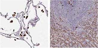
IHC staining of human lung tissue using the Anti-CD68 antibody (HPA048982) shows strong cytoplasmic positivity in macrophages and in hematopoietic tissues, such as spleen.

DTL

DTL

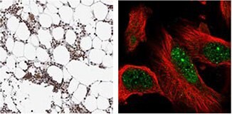
IHC staining of human bone marrow using the Anti-DTL antibody (HPA028016) shows strong nuclear positivity in bone marrow poietic cells. By ICC-IF, staining of nucleus in U-251 MG cells is detected.

GSTM5

GSTM5

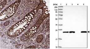
The Anti-GSTM5 antibody (HPA048652) shows cytoplasmic positivity in glandular cells in human rectum by IHC and in WB, the antibody detects a band of predicted size in cell lysates of RT-4, U-251 MG, as well as in liver tissue lysate.

* WB both in human and rodent samples

1.
Gill RM, Gabor TV, Couzens AL, Scheid MP. 2013. The MYC-Associated Protein CDCA7 Is Phosphorylated by AKT To Regulate MYC-Dependent Apoptosis and Transformation. Mol. Cell. Biol.. 33(3):498-513. https://doi.org/10.1128/mcb.00276-12
2.
Shubbar E, Kovács A, Hajizadeh S, Parris TZ, Nemes S, Gunnarsdóttir K, Einbeigi Z, Karlsson P, Helou K. 2013. Elevated cyclin B2 expression in invasive breast carcinoma is associated with unfavorable clinical outcome. BMC Cancer. 13(1): https://doi.org/10.1186/1471-2407-13-1
3.
Karaayvaz M, Pal T, Song B, Zhang C, Georgakopoulos P, Mehmood S, Burke S, Shroyer K, Ju J. 2011. Prognostic Significance of miR-215 in Colon Cancer. Clinical Colorectal Cancer. 10(4):340-347. https://doi.org/10.1016/j.clcc.2011.06.002
4.
Bozóky B, Savchenko A, Csermely P, Korcsmáros T, Dúl Z, Pontén F, Székely L, Klein G. 2013. Novel signatures of cancer-associated fibroblasts. Int. J. Cancer. 133(2):286-293. https://doi.org/10.1002/ijc.28035
5.
Rognum IJ, Haynes RL, Vege ?, Yang M, Rognum TO, Kinney HC. 2009. Interleukin-6 and the serotonergic system of the medulla oblongata in the sudden infant death syndrome. Acta Neuropathol. 118(4):519-530. https://doi.org/10.1007/s00401-009-0535-y

MMP9

mmp9-1

IHC staining of human lung tissue using the Anti-MMP9 antibody (HPA001238) shows strong nuclear positivity in macrophages and in bone marrow poietic cells in bone marrow tissue.

mmp9-2

Monclonal Anti-MMP9 antibodies show strong cytoplasmic positivity in a subset of lymphoid cells in duodenum (AMAb90805) and in human tonsil tissue (AMAb90804).

LYRIC/MTDH

lyric-mtdh-1

IHC staining using the Anti-MTDH antibody (HPA010932) shows strong cytoplasmic positivity in neuronal cells in human cerebral cortex tissue. In ICC-IF in A-431 cell line, the antibody stains endoplasmic reticulum.

lyric-mtdh-2

IHC staining using the monclonal Anti-MTDH antibody (AMAb90762) shows strong cytoplasmic reactivity in tumor cells from breast and colorectal cancer samples.

P5C Dehydrogenase/ALDH4A1

p5c-dehydrogenase-aldh4a1

IHC staining using the Anti- ALDH4A1 antibody (HPA006401) shows strong cytoplasmic positivity with granular pattern in human kidney and liver tissues.

* WB both in human and rodent samples

1.
Pohler E, Mamai O, Hirst J, Zamiri M, Horn H, Nomura T, Irvine AD, Moran B, Wilson NJ, Smith FJD, et al. 2012. Haploinsufficiency for AAGAB causes clinically heterogeneous forms of punctate palmoplantar keratoderma. Nat Genet. 44(11):1272-1276. https://doi.org/10.1038/ng.2444
2.
Roca H, Craig MJ, Ying C, Varsos ZS, Czarnieski P, Alva AS, Hernandez J, Fuller D, Daignault S, Healy PN, et al. 2011. IL-4 induces proliferation in prostate cancer PC3?cells under nutrient-depletion stress through the activation of the JNK-pathway and survivin upregulation. J. Cell. Biochem..n/a-n/a. https://doi.org/10.1002/jcb.24025
3.
Friedman JS, Chang B, Krauth DS, Lopez I, Waseem NH, Hurd RE, Feathers KL, Branham KE, Shaw M, Thomas GE, et al. 2010. Loss of lysophosphatidylcholine acyltransferase 1 leads to photoreceptor degeneration in rd11 mice. Proceedings of the National Academy of Sciences. 107(35):15523-15528. https://doi.org/10.1073/pnas.1002897107
4.
Nohata N, Hanazawa T, Kikkawa N, Mutallip M, Sakurai D, Fujimura L, Kawakami K, Chiyomaru T, Yoshino H, Enokida H, et al. 2011. Tumor suppressive microRNA-375 regulates oncogene AEG-1/MTDH in head and neck squamous cell carcinoma (HNSCC). J Hum Genet. 56(8):595-601. https://doi.org/10.1038/jhg.2011.66
5.
LIU B, WU Y, PENG D. 2013. Astrocyte elevated gene-1 regulates osteosarcoma cell invasion and chemoresistance via endothelin-1/endothelin A receptor signaling. 5(2):505-510. https://doi.org/10.3892/ol.2012.1056
6.
Lorenzen JM, Martino F, Scheffner I, Bröcker V, Leitolf H, Haller H, Gwinner W. Fetuin, Matrix-Gla Protein and Osteopontin in Calcification of Renal Allografts. PLoS ONE. 7(12):e52039. https://doi.org/10.1371/journal.pone.0052039

PITRM1/MP1

pitrm1-mp1

The Anti- PITRM1 antibody (HPA006753) shows strong cytoplasmic positivity in myocytes in human heart muscle using IHC. ICC-IF staining of human cell line U-251 MG shows positivity in mitochondria.

PRC1

prc1

IHC staining of human testis tissue using the Anti-PRC1 antibody (HPA034521) shows strong nuclear positivity in cells of seminiferus ducts. ICC-IF shows staining of nucleus, plasma membrane and microtubules in A-431 cells.

SCOT/OXCT1

scot-oxct1

IHC staining of human heart muscle and kidney by Anti-OXCT1 antibody (HPA012047) shows strong cytoplasmic positivity in myocytes and cells in tubules, respectively. ICC-IF shows staining of mitochondria in A431 cells.

* WB both in human and rodent samples

1.
Pereira CBL, Leal MF, de Souza CRT, Montenegro RC, Rey JA, Carvalho AA, Assumpção PP, Khayat AS, Pinto GR, Demachki S, et al. Prognostic and Predictive Significance of MYC and KRAS Alterations in Breast Cancer from Women Treated with Neoadjuvant Chemotherapy. PLoS ONE. 8(3):e60576. https://doi.org/10.1371/journal.pone.0060576
2.
Chang HT, Olson L, Schwartz KA. 2013. Ketolytic and glycolytic enzymatic expression profiles in malignant gliomas: implication for ketogenic diet therapy. Nutrition & Metabolism. 10(1):47. https://doi.org/10.1186/1743-7075-10-47
3.
Zibert JR, Wallbrecht K, Schön M, Mir LM, Jacobsen GK, Trochon-Joseph V, Bouquet C, Villadsen LS, Cadossi R, Skov L, et al. 2011. Halting angiogenesis by non-viral somatic gene therapy alleviates psoriasis and murine psoriasiform skin lesions. J. Clin. Invest.. 121(1):410-421. https://doi.org/10.1172/jci41295

Antibodies Identified in the Human Protein Atlas

Showing Differential IHC Staining Patterns in Breast Cancer Samples

anti-klhl26-antibody

IHC analysis using Anti-KLHL26 antibody (HPA023074) shows a varying membranous/cytoplasmic staining pattern in breast tumor samples from different patients.

anti-acsf2-antibody

The Anti-ACSF2 (HPA024693) antibody shows granular cytoplasmic positivity in breast tumor cells from different patients varying from strong to negative.

anti-gcm1-antibody

The Anti-GCM1 (HPA011343) antibody shows membranous positivity in breast tumor cells while normal breast tissue is negative.

anti-agr3-antibody

The Anti-AGR3 (HPA053942) antibody shows strong cytoplasmic positivity in 11/12 breast cancer patients, while 1 patient is completely negative.

* WB both in human and rodent samples

1.
Ngan E, Diamond B. 2012. LPP is Required for TGF-Beta Induced Motility and Invasion of Neu/ErbB-2 Expressing Breast Cancer Cells. https://doi.org/10.21236/ada568114
2.
Camilleri M, Carlson P, Zinsmeister AR, McKinzie S, Busciglio I, Burton D, Zucchelli M, D'Amato M. 2010. Neuropeptide S Receptor Induces Neuropeptide Expression and Associates With Intermediate Phenotypes of Functional Gastrointestinal Disorders. Gastroenterology. 138(1):98-107.e4. https://doi.org/10.1053/j.gastro.2009.08.051
3.
Bozóky B, Savchenko A, Csermely P, Korcsmáros T, Dúl Z, Pontén F, Székely L, Klein G. 2013. Novel signatures of cancer-associated fibroblasts. Int. J. Cancer. 133(2):286-293. https://doi.org/10.1002/ijc.28035
4.
Stro?mberg S, Agnarsdo?ttir M, Magnusson K, Rexhepaj E, Bolander Å, Lundberg E, Asplund A, Ryan D, Rafferty M, Gallagher WM, et al. 2009. Selective Expression of Syntaxin-7 Protein in Benign Melanocytes and Malignant Melanoma. J. Proteome Res.. 8(4):1639-1646. https://doi.org/10.1021/pr800745e

Finding Cancer Biomarkers

Breast Cancer

Breast cancer is the second most common cancer and by far the most frequent cancer among women. The incidence of breast cancer is increasing steadily, but without a corresponding increase in mortality. If detected at an early stage, the prognosis is relatively good for a patient living in a developed country, with a general five-year survival rate of approximately 85%.

Breast Cancer and Treatment

Cancer, though often denoted as a singular disease, is truly a multitude of diseases. This understanding has evolved over the years, but many patients are not receiving optimal treatment for their disease. For cancer patients to receive a more individualized treatment, there is still a need for new and better ways to stratify patients. The classical prognostic factors such as stage and grade of the tumor are insufficient for a correct estimation of patient prognosis. Additional information from cancer biomarkers promise to substantially improve this estimation, ultimately leading to a more individualized treatment, thus avoiding both under and overtreatment of patients.

The primary curative treatment for breast cancer patients is surgery, often in combination with adjuvant therapy. However, adjuvant therapy is associated with substantial costs and sometimes severe side effects, and physicians have identified reduction of overtreatment as the major clinical need in breast cancer treatment today. Thus, the stratification of patients into different prognostic categories is of great importance as it may aid physicians in selecting the most appropriate treatment for a given patient.

The majority of breast cancers are hormone receptor responsive, i.e., express the estrogen receptor (ER) and/or the progesteron receptor (PR). Patients with tumors expressing these receptors may receive adjuvant endocrine treatment, such as tamoxifen.

Breast cancers may also express the HER2 protein (human epidermal growth factor receptor 2), and patients with tumors expressing this protein may receive adjuvant therapy with trastuzumab.

Adjuvant treatment may also consist of chemotherapy or radiation therapy.

RBM3

The RNA-binding motif protein 3 (RBM3) is an RNA- and DNAbinding protein, whose function has not been fully elucidated. It has been shown that the protein is expressed as an early event in mild hypothermia, and also in other conditions relating to cellular stress, such as glucose deprivation and hypoxia1. During stress, RBM3 is thought to protect the cells by aiding in maintenance of protein synthesis needed for survival1. Recently, it has also been shown that RBM3 attenuates stem cell-like properties in prostate cancer cells2.

RBM3 was identified via the Human Protein Atlas (HPA) as a potential oncology biomarker through the differential expression pattern present in several cancers investigated as part of the HPA project (proteinatlas.org)3,4.

The IHC analysis using the Anti-RBM3 antibody HPA003624 showed a weak expression pattern in normal breast tissue, but a stratified pattern in breast cancer tissue (Figure 1). Researchers further investigated the expression in larger breast cancer cohorts and the expression of RBM3 was shown to be associated with a prolonged survival5.

1.
Ehlén Å. 2011. Te Role of RNA-Binding Motif 3 in Epithelial Ovarian Cancer: A Biomarker Discovery Approach .
2.
Zeng Y, Wodzenski D, Gao D, Shiraishi T, Terada N, Li Y, Vander Griend DJ, Luo J, Kong C, Getzenberg RH, et al. 2013. Stress-Response Protein RBM3 Attenuates the Stem-like Properties of Prostate Cancer Cells by Interfering with CD44 Variant Splicing. Cancer Res. 73(13):4123-4133. https://doi.org/10.1158/0008-5472.can-12-1343
3.
Berglund L, Björling E, Oksvold P, Fagerberg L, Asplund A, Al-Khalili Szigyarto C, Persson A, Ottosson J, Wernérus H, Nilsson P, et al. 2008. A Genecentric Human Protein Atlas for Expression Profiles Based on Antibodies. Mol Cell Proteomics. 7(10):2019-2027. https://doi.org/10.1074/mcp.r800013-mcp200
4.
Uhlen M, Oksvold P, Fagerberg L, Lundberg E, Jonasson K, Forsberg M, Zwahlen M, Kampf C, Wester K, Hober S, et al. 2010. Towards a knowledge-based Human Protein Atlas. Nat Biotechnol. 28(12):1248-1250. https://doi.org/10.1038/nbt1210-1248
5.
Jögi A, Brennan DJ, Rydén L, Magnusson K, Fernö M, Stål O, Borgquist S, Uhlen M, Landberg G, Påhlman S, et al. 2009. Nuclear expression of the RNA-binding protein RBM3 is associated with an improved clinical outcome in breast cancer. Mod Pathol. 22(12):1564-1574. https://doi.org/10.1038/modpathol.2009.124
RBM3 Antibody

Figure 1. Immunohistochemical analysis using the Anti-RBM3 antibody (HPA003624) shows weak expression in normal breast tissue (A) and differential expression, varying from weak to strong in tumor breast samples (B, C).

RBM3 as a Prognostic Biomarker in Breast Cancer

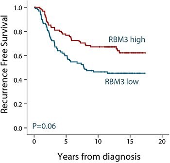
After identification of RBM3 as a potential prognostic biomarker, researchers further investigated the RBM3 protein expression in larger breast cancer cohorts1. In a cohort of 500 premenopausal women with stage II invasive breast cancer, RBM3 expression was found to be associated with small, low-grade, estrogen receptor (ER)-positive tumors. When analyzing the subset of ER-positive patients, RBM3 was an independent predictor of recurrence free survival (RFS). As shown in Figure 2, patients with tumors expressing high levels of the RBM3 protein have an improved survival compared to patients with tumors expressing low levels of RBM3.

RBM3 protein expression has further been analyzed in many different patient cohorts from various forms of cancer. Levels of RBM3 expression was found to have a significant connection to patient survival in breast1, colon2, ovarian3,4, testicular, urothelial5, and prostate6 cancer as well as in malignant melanoma7.

In conclusion, RBM3 is a marker of good prognosis in breast cancer as well as in several other cancers.
kaplan-meier-survival-analysis

Figure 2. Kaplan-Meier (survival) analysis of recurrence free survival (RFS) according to RBM3 expression for ER-positive breast cancer patients. Patients were split into two groups based on high and low RBM3 expression.

RBM3 Antibodies

There are two Anti-RBM3 antibodies in Atlas Antibodies' product catalog; the polyclonal HPA003624 and the PrecisA Monoclonal AMAb90655. The monoclonal Anti-RBM3 antibody AMAb90655 has shown excellent specificity in Western blot analysis of human cell lines, and is routinely used for staining of formalin fixed paraffin embedded tissue in IHC (Figure 3).

RBM3 Antibodies

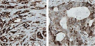
Figure 3. Immunohistochemical analysis of RBM3 expression in breast cancer (left) and prostate cancer (right) using AMAb90655 shows nuclear positivity in tumor cells. The WB image shows an expected band of 17 kDa in human cell line RT4 lysate using AMAb90655.

References

1.
Jögi A, Brennan DJ, Rydén L, Magnusson K, Fernö M, Stål O, Borgquist S, Uhlen M, Landberg G, Påhlman S, et al. 2009. Nuclear expression of the RNA-binding protein RBM3 is associated with an improved clinical outcome in breast cancer. Mod Pathol. 22(12):1564-1574. https://doi.org/10.1038/modpathol.2009.124
2.
Hjelm B, Brennan DJ, Zendehrokh N, Eberhard J, Nodin B, Gaber A, Pontén F, Johannesson H, Smaragdi K, Frantz C, et al. 2011. High nuclear RBM3 expression is associated with an improved prognosis in colorectal cancer. Prot. Clin. Appl.. 5(11-12):624-635. https://doi.org/10.1002/prca.201100020
3.
Ehlén Å, Brennan DJ, Nodin B, O'Connor DP, Eberhard J, Alvarado-Kristensson M, Jeffrey IB, Manjer J, Brändstedt J, Uhlén M, et al. 2010. Expression of the RNA-binding protein RBM3 is associated with a favourable prognosis and cisplatin sensitivity in epithelial ovarian cancer. Journal of Translational Medicine. 8(1):78. https://doi.org/10.1186/1479-5876-8-78
4.
Ehlén Õ, Nodin B, Rexhepaj E, Brändstedt J, Uhlén M, Alvarado-Kristensson M, Pontén F, Brennan DJ, Jirström K. 2011. RBM3-Regulated Genes Promote DNA Integrity and Affect Clinical Outcome in Epithelial Ovarian Cancer. Translational Oncology. 4(4):212-IN1. https://doi.org/10.1593/tlo.11106
5.
Boman K, Segersten U, Ahlgren G, Eberhard J, Uhlén M, Jirström K, Malmström P. 2013. Decreased expression of RNA-binding motif protein 3 correlates with tumour progression and poor prognosis in urothelial bladder cancer. BMC Urol. 13(1): https://doi.org/10.1186/1471-2490-13-17
6.
Jonsson L, Gaber A, Ulmert D, Uhlén M, Bjartell A, Jirström K. 2011. High RBM3 expression in prostate cancer independently predicts a reduced risk of biochemical recurrence and disease progression. Diagn Pathol. 6(1): https://doi.org/10.1186/1746-1596-6-91
7.
Jonsson L, Bergman J, Nodin B, Manjer J, Pontén F, Uhlén M, Jirström K. 2011. Low RBM3 protein expression correlates with tumour progression and poor prognosis in malignant melanoma: An analysis of 215 cases from the Malmö Diet and Cancer Study. Journal of Translational Medicine. 9(1):114. https://doi.org/10.1186/1479-5876-9-114

Granulin

Granulins are a family of secreted, glycosylated peptides that are cleaved from a single precursor protein. Cleavage of the signal peptide produces mature granulin which can be further cleaved into a variety of active peptides. These cleavage products are named granulin A, granulin B, granulin C, etc. Both the peptides and intact granulin protein regulate cell growth. Different members of the granulin protein family may act as inhibitors, stimulators, or have dual actions on cell growth. Granulin family members are important in normal development, wound healing, and tumorigenesis [provided by RefSeq, Jul 2008].

In a paper by Elkabets et al, the role of GRN expression in responding tumor instigation was investigated by studying recrution of GRNexpressing bone marrow cells into responding tumors in mice1. Certain tumors can foster the growth of other tumors or metastatic cells located at distant anatomical sites, which is referred to as tumor instigation. In this study, rigorously growing human breast carcinoma cells were implanted in mice and it was shown that these cells stimulated the outgrowth of otherwise poorly tumorigenic, indolent transformed cells. GRN was identified as the most upregulated gene in the instigating bone marrow cells. The GRN expressing cells induced resident fibroblasts to express genes that promoted malignant tumor progression. It was speculated whether anticancer therapies might involve targeting GRN, or the activated GRN expressing cells, and thereby disrupting these cell lines of communication that promote cancer progression.

By using the Anti-GRN antibody HPA028747 in the analysis of tumor tissues from a cohort of breast cancer patients, high GRN expression was shown to correlate with the most aggressive triple-negative, basal-like tumor subtype and reduced patient survival (Figure 4).

References

1.
Elkabets M, Gifford AM, Scheel C, Nilsson B, Reinhardt F, Bray M, Carpenter AE, Jirström K, Magnusson K, Ebert BL, et al. 2011. Human tumors instigate granulin-expressing hematopoietic cells that promote malignancy by activating stromal fibroblasts in mice. J. Clin. Invest.. 121(2):784-799. https://doi.org/10.1172/jci43757
grn-expression

Figure 4. GRN expression was shown to correlate with aggressive tumor subtypes and reduced survival of breast cancer patients using antibody HPA028747. The diagram to the left shows percentage of tumors in each category (Triple-Negative [TN]/ basal or nonbasal) that show high GRN positivity and the Kaplan-Meier analysis to the right shows correlation between GRN-positive (green) or GRN-negative (blue) expression and survival.

Granulin Antibodies

In Atlas Antibodies´ product catalog, there are two polyclonal Anti-GRN antibodies; HPA008763 and HPA028747.

ihc-staining-of-human-pancreas-tissue

IHC staining of human pancreas tissue using the Anti-GRN antibody (HPA008763) shows strong cytoplasmic positivity in exocrine glandular cells. ICC-IF shows positivity in vesicles in A-431 cells.

ihc-analysis-using-the-anti-grn-antibody

IHC analysis using the Anti-GRN antibody HPA028747 shows strong cytoplasmic positivity in normal duodenum tissue in glanduclar cells and vesicle positivity in U-251 MG cells.

Anillin

Anillin is an actin binding protein that is a subunit of microfilaments, one of the cytoskeleton components. Anillin is expressed in most cells and is involved in basic cell functions, e.g. motility, division and signaling. Studies of anillin expression have shown that it is over expressed in several human tumors.

Anillin as a Treatment Predictive Prognostic Biomarker in Breast Cancer

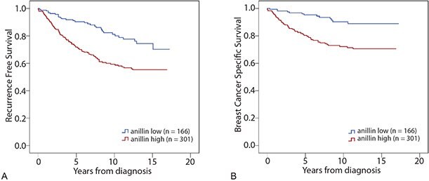
Anillin expression was analyzed in a patient cohort consisting of 467 samples from patients diagnosed with breast cancer, using the Anti-ANLN antibody HPA005680. Patients with tumors expressing high levels of anillin had a reduced recurrence free survival (RFS) compared to patients with tumors expressing low levels of anillin (Figure 5A). The same association between anillin expression and reduced survival could be seen when analyzing breast cancer specific survival (BCSS, Figure 5B). In a study by O´Leary et al, the prognostic impact of anillin was confirmed by Cox regression analysis. High anillin expression was associated with reduced BCSS and RFS in univariate- as well as in multivariate analysis, adjusted for tumor size and grade, age at diagnosis, nodal-, ER-, PR-, HER2-, and Ki67 status.

In conclusion, anillin is a marker for poor prognosis in breast cancer.
kaplan-meier-survival-analysis-2

Figure 5. Kaplan-Meier (survival) analysis of recurrence free- (A) and breast cancer specific survival (B) according to aniliin expression for breast cancer patients. Patients were split into two groups based on high and low anillin expression.

References

1.
O´Leary PC, Penny SA, Dolan RT, Kelly CM, Madden SF, Rexhepaj E, Brennan DJ, McCann AH, Pontén F, Uhlén M, et al. 2013. Systematic antibody generation and validation via tissue microarray technology leading to identification of a novel protein prognostic panel in breast cancer. BMC Cancer. 13(1): https://doi.org/10.1186/1471-2407-13-175

Anillin Antibodies

There are three Anti-ANLN antibodies in Atlas Antibodies product catalog; the monoclonals AMAb90660 and AMAb90662, and the polyclonal HPA005680.

anillin-antibodies
anti-anln-antibody

The Anti-ANLN antibody (HPA005680) shows strong nuclear positivity in cells in seminiferous ducts in human testis by IHC. In ICC-IF, nuclei (but not nucleoli) of A-431 cells stain positively and in WB, the antibody detects a band of predicted size in cell lysates of RT-4 and U-251 MG.
anti-anln-antibody-2

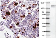
Anti-ANLN antibody AMAb90660 shows strong nuclear immunoreactivity in a subset of tumour cells in lung adenocarcinoma and a band of predicted size in human cell line U-251 MG.

anti-anln-antibody-3

AMAb90662 Anti-ANLN antibody shows strong nuclear immunoreactivity in a subset of tumor cells in colorectal cancer and a band of predicted size in human U-251 MG cells.

Zaloguj się, aby kontynuować

Zaloguj się lub utwórz konto, aby kontynuować.

Nie masz konta użytkownika?