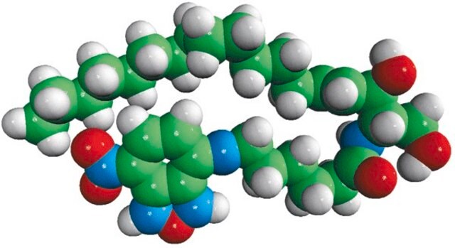
SCT105
BioTracker NucView® 530 Red Caspase-3 Dye (PBS)
Live cell imaging apoptosis dye for caspase-3/7 enzyme activity used to detect programmed cell death using fluorescence microscopy or flow cytometry applications.
Synonym(s):
Live cell caspase-3 apoptosis probe, Live cell imaging probe
Sign Into View Organizational & Contract Pricing
All Photos(1)
About This Item
UNSPSC Code:
12352207
NACRES:
NA.47
Recommended Products
technique(s)
cell based assay: suitable
detection method
fluorometric
shipped in
ambient
General description
Caspases are a family of protease enzymes that play essential roles in programmed cell death, termed apoptosis. Caspase-3 is a caspase protein that interacts with caspase-8 and caspase-9 and sequential activation of plays a central role in the execution-phase of cellular apoptosis.
The BioTracker NucView 530 Red Caspase-3 Dye provides a convenient live cell tool for profiling apoptotic cell populations based on caspase-3/7 activity using either fluorescence microscopy or flow cytometry. In contrast to other fluorogenic caspase substrates or fluorescent caspase inhibitor based (FLICA) assays, the BioTracker NucView 530 Red Caspase-3 Dye can be used to detect caspase-3/7 activity within individual intact cells without inhibiting apoptosis progression. The substrate consists of a fluorogenic DNA dye coupled to the caspase-3/7 DEVD recognition sequence. The substrate, which is initially non-fluorescent, penetrates the plasma membrane and enters the cytoplasm. In apoptotic cells, caspase-3/7 cleaves the substrate, releasing the high-affinity DNA dye, which migrates to the cell nucleus and stains DNA with bright red fluorescence. Thus, NucView 530 Caspase-3 Substrate is bifunctional, allowing detection of caspase-3/7 activity and visualization of morphological changes in the nucleus during apoptosis. The dye is formaldehyde-fixable and compatible with subsequent immunostaining. The dye is offered in DMSO and PBS (phosphate-buffered saline) formulations. The substrate in PBS is formulated for use in cells that are sensitive to DMSO toxicity.
NucView 530 Caspase-3 Substrate stains apoptotic cell nuclei with orange fluorescence, for detection in the Cy3 channel by fluorescence microscopy or the PE channel by flow cytometry. Note that when excited by the 488nm laser line, NucView 530 also fluoresces in the FITC channel, and therefore cannot be analyzed together with green probes by flow cytometry.
Application
Live cell fluorescent imaging
Live cell imaging apoptosis dye for caspase-3/7 enzyme activity used to detect programmed cell death using fluorescence microscopy or flow cytometry applications.
Research Category
Cell Imaging
Cell Imaging
Research Sub Category
Live Cell Dye
Live Cell Dye
Quality
Absorbance Spectrum: 523 +/- 5
TLC: >95%
TLC: >95%
Physical form
Liquid
Storage and Stability
Store BioTracker NucView 530 Red Caspase-3 Dye, 1mM in PBS at -20ºC. Protect From Light.
Note: Centrifuge vial briefly to collect contents at bottom of vial before opening.
Note: Centrifuge vial briefly to collect contents at bottom of vial before opening.
Legal Information
NucView is a registered trademark of Biotium Inc.
Disclaimer
Unless otherwise stated in our catalog or other company documentation accompanying the product(s), our products are intended for research use only and are not to be used for any other purpose, which includes but is not limited to, unauthorized commercial uses, in vitro diagnostic uses, ex vivo or in vivo therapeutic uses or any type of consumption or application to humans or animals.
Storage Class Code
12 - Non Combustible Liquids
WGK
WGK 1
Certificates of Analysis (COA)
Search for Certificates of Analysis (COA) by entering the products Lot/Batch Number. Lot and Batch Numbers can be found on a product’s label following the words ‘Lot’ or ‘Batch’.
Already Own This Product?
Find documentation for the products that you have recently purchased in the Document Library.
Our team of scientists has experience in all areas of research including Life Science, Material Science, Chemical Synthesis, Chromatography, Analytical and many others.
Contact Technical Service


