Saltar al contenido
Merck
Todas las fotos(2)

Key Documents

MABE340-AF555

Sigma-Aldrich

Anti-MDM2 Antibody, clone IF2, Alexa Fluor 555 Conjugate

clone IF2, from mouse, ALEXA FLUOR 555

Sinónimos:

E3 ubiquitin-protein ligase Mdm2, Double minute 2 protein, Hdm2, Oncoprotein Mdm2, p53-binding protein Mdm2

Iniciar sesiónpara Ver la Fijación de precios por contrato y de la organización

Seleccione un Tamaño


Seleccione un Tamaño

Cambiar Vistas

About This Item

UNSPSC Code:
12352203
eCl@ss:
32160702
NACRES:
NA.45

biological source

mouse

Quality Level

conjugate

ALEXA FLUOR 555

antibody form

purified antibody

antibody product type

primary antibodies

clone

IF2, monoclonal

species reactivity

human

technique(s)

immunocytochemistry: suitable

isotype

IgG2b

NCBI accession no.

General description

E3 ubiquitin-protein ligase Mdm2 (UniProt Q00987; also known as Double minute 2 protein, Hdm2, Oncoprotein Mdm2, p53-binding protein Mdm2) is encoded by the MDM2 gene (Gene ID 4193) in human. MDM2 is an E3 ubiquitin ligase that is most widely studied for its role in regulating the p53 tumor suppressor. MDM2 antagonizes p53 transcriptional activity by binding to and blocking p53 N-terminal trans-activation domain. MDM2 can also monoubiquitinate p53, thereby promoting p53 cytoplasmic localization to prevent p53 nuclear funtion. In addition, MDM2, when in complex with p300, also mediates p53 polyubiquitination, resulting in p53 proteosomal degradation. MDM2 overexpression is shown to inhibit DNA double-strand break repair mediated through its interaction with Nbs1 in a p53-independent manner.

Specificity

Clone IF2 recognizes the ~85/90 kDa (apparent MW) MDM2, as well as isoforms of ~57/60 kDa and ~74/76 kDa by immunoblotting using the unconjugated antibody (Cat. Nos. MABE340 & OP46).

Immunogen

Epitope: Within amino acids 26-169.
Recombinant human MDM2.

Application

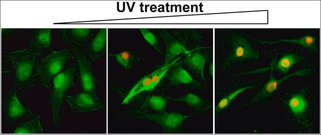
Immunocytochemistry Analysis: A 1:100 dilution from a representative lot detected MDM2 in HeLa cells.
The unconjugated antibody (Cat. Nos. MABE340 & OP46) is shown to be suitable also for immunohistochemistry and Western blotting applications.
Research Category
Epigenetics & Nuclear Function
Research Sub Category
Apoptosis - Additional
This Anti-MDM2 Antibody, clone IF2, Alexa Fluor 555 Conjugate is validated for use in Immunocytochemistry for the detection of MDM2.

Quality

Evaluated by Immunocytochemistry in HepG2 cells.

Immunocytochemistry Analysis: A 1:100 dilution of this antibody detected MDM2 in HepG2 cells.

Target description

~85 kDa observed.

Physical form

Protein G purified.
Purified mouse monoclonal IgG2b antibody conjugate in PBS with 15 mg/mL BSA and 0.1 % sodium azide.

Storage and Stability

Stable for 1 year at 2-8°C from date of receipt.

Other Notes

Concentration: Please refer to lot specific datasheet.

Legal Information

ALEXA FLUOR is a trademark of Life Technologies

Disclaimer

Unless otherwise stated in our catalog or other company documentation accompanying the product(s), our products are intended for research use only and are not to be used for any other purpose, which includes but is not limited to, unauthorized commercial uses, in vitro diagnostic uses, ex vivo or in vivo therapeutic uses or any type of consumption or application to humans or animals.

¿No encuentra el producto adecuado?  

Pruebe nuestro Herramienta de selección de productos.

Storage Class

12 - Non Combustible Liquids

wgk_germany

WGK 2


  • Certificados de análisis (COA)

    Busque Certificados de análisis (COA) introduciendo el número de lote del producto. Los números de lote se encuentran en la etiqueta del producto después de las palabras «Lot» o «Batch»

    ¿Ya tiene este producto?

    Encuentre la documentación para los productos que ha comprado recientemente en la Biblioteca de documentos.

    Visite la Librería de documentos

    Nuestro equipo de científicos tiene experiencia en todas las áreas de investigación: Ciencias de la vida, Ciencia de los materiales, Síntesis química, Cromatografía, Analítica y muchas otras.

    Póngase en contacto con el Servicio técnico