Skip to Content
Merck
All Photos(5)

Key Documents

05-649

Sigma-Aldrich

Anti-Chk2 Antibody, clone 7

clone 7, Upstate®, from mouse

Synonym(s):

CHK2 (checkpoint, S.pombe) homolog, CHK2 checkpoint homolog (S. pombe), checkpoint-like protein CHK2, protein kinase CHK2, serine/threonine-protein kinase CHK2

Sign Into View Organizational & Contract Pricing


About This Item

UNSPSC Code:
12352203
eCl@ss:
32160702
NACRES:
NA.41

biological source

mouse

Quality Level

antibody form

purified immunoglobulin

antibody product type

primary antibodies

clone

7, monoclonal

species reactivity

mouse, human

manufacturer/tradename

Upstate®

technique(s)

immunocytochemistry: suitable
immunoprecipitation (IP): suitable
western blot: suitable

isotype

IgG1

NCBI accession no.

UniProt accession no.

shipped in

dry ice

target post-translational modification

unmodified

Gene Information

human ... CHEK2(11200)

General description

The p53 tumor-suppressor gene integrates numerous signals that control cell life and death. Several novel molecules involved in p53 signaling, including Chk2, p53R2, p53AIP1, Noxa, PIDD, and PID/MTA2, were recently discovered. The checkpoint kinase Chk2 is the mammalian homologue of yeast Cds1/Rad53. In response to DNA damage, the checkpoint kinase ATM phosphorylates and activates Chk2, which in turn directly phosphorylates and activates p53. Chk2 serves as ATM downstream effector to mediate activation of p53. Chk2 also phosphorylates and activates BRCA1, the product of a tumor suppressor gene that is mutated in breast and ovarian cancer.

Specificity

Chk2
Others not tested.
Predicted to cross-react with rat based on sequence homology.

Immunogen

Mixture of two GST fusion proteins corresponding to residues 1-223 and 217-543 of human Chk2

Application

Anti-Chk2 Antibody, clone 7 is a mouse monoclonal antibody for detection of Chk2 also known as CHK2 checkpoint homolog (S. pombe), serine/threonine-protein kinase CHK2 & has been validated in WB, IP, ICC.
Immunoprecipitation: A previous lot of this antibody has been reported by an independent laboratory to immunoprecipitate Chk2 from HeLa cells.

Immunocytochemistry: A previous lot of this antibody has been reported by an independent laboratory to detect Chk2 in HeLa cells.

Immunocytochemistry Analysis: A 1:1000 dilution from a representative lot detected Chk2 in HeLa cells.
Research Category
Epigenetics & Nuclear Function
Research Sub Category
Cell Cycle, DNA Replication & Repair

Quality

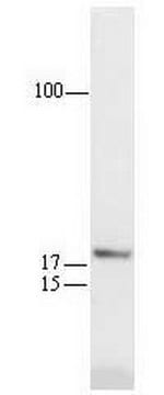
Routinely evaluated by western blot on RIPA lysates from Jurkat cells, HeLa, 3T3/A31, or A431 cells.

Western Blot Analysis: 0.1-1 μg/mL of this lot detected Chk2 in RIPA lysates from Jurkat cells. A previous lot detected Chk2 in lysates from HeLa, 3T3/A31, and A431 cells.

Target description

67 kDa

Linkage

Replaces: MABE491

Physical form

Format: Purified
Protein G Chromatography
Protein G purified mouse IgG1 buffer containing 0.07 M Tris-glycine, pH 7.4, 0.105 M NaCl, 0.035% sodium azide and 30% glycerol. Liquid at -20°C.

Storage and Stability

Stable for 1 year at from date of receipt.

Analysis Note

Control
Positive Antigen Control: Catalog #12-303, Jurkat cell lysate.

Other Notes

Concentration: Please refer to the Certificate of Analysis for the lot-specific concentration.

Legal Information

UPSTATE is a registered trademark of Merck KGaA, Darmstadt, Germany

Disclaimer

Unless otherwise stated in our catalog or other company documentation accompanying the product(s), our products are intended for research use only and are not to be used for any other purpose, which includes but is not limited to, unauthorized commercial uses, in vitro diagnostic uses, ex vivo or in vivo therapeutic uses or any type of consumption or application to humans or animals.

Not finding the right product?  

Try our Product Selector Tool.

Storage Class Code

10 - Combustible liquids

WGK

WGK 1


Certificates of Analysis (COA)

Search for Certificates of Analysis (COA) by entering the products Lot/Batch Number. Lot and Batch Numbers can be found on a product’s label following the words ‘Lot’ or ‘Batch’.

Already Own This Product?

Find documentation for the products that you have recently purchased in the Document Library.

Visit the Document Library

Jeffrey M Sifford et al.
Journal of virology, 90(5), 2571-2585 (2015-12-18)
Tumor suppressor p53 is activated in response to numerous cellular stresses, including viral infection. However, whether murine gammaherpesvirus 68 (MHV68) provokes p53 during the lytic replication cycle has not been extensively evaluated. Here, we demonstrate that MHV68 lytic infection induces
X Wu et al.
The Journal of biological chemistry, 276(4), 2971-2974 (2000-10-29)
The integrity of the DNA damage response pathway is essential for prevention of neoplastic transformation. Several proteins involved in this pathway including p53, BRCA1, and ATM are frequently mutated in human cancer. Checkpoint kinase 2 (Chk2) is a DNA damage-activated
Richard O Adeyemi et al.
PLoS pathogens, 6(10), e1001141-e1001141 (2010-10-16)
Infection by DNA viruses can elicit DNA damage responses (DDRs) in host cells. In some cases the DDR presents a block to viral replication that must be overcome, and in other cases the infecting agent exploits the DDR to facilitate
Kazumasa Yoshida et al.
Journal of cell science, 123(Pt 2), 225-235 (2010-01-06)
CDC6, a replication licensing protein, is partially exported to the cytoplasm in human cells through phosphorylation by Cdk during S phase, but a significant proportion remains in the nucleus. We report here that human CDC6 physically interacts with ATR, a
Víctor García et al.
Oncotarget, 7(4), 4490-4506 (2015-12-20)
Galiellalactone (GL) is a fungal metabolite that presents antitumor activities on prostate cancer in vitro and in vivo. In this study we show that GL induced cell cycle arrest in G2/M phase, caspase-dependent apoptosis and also affected the microtubule organization

Our team of scientists has experience in all areas of research including Life Science, Material Science, Chemical Synthesis, Chromatography, Analytical and many others.

Contact Technical Service