Skip to Content
Merck
All Photos(5)

Documents

06-570

Sigma-Aldrich

Anti-phospho-Histone H3 (Ser10) Antibody, Mitosis Marker

Upstate®, from rabbit

Synonym(s):

H3 histone, family 3A, H3S10P, Histone H3 (phospho S10), H3 histone, family 3A, H3 histone, family 3B, H3 histone, family 3B (H3.3B)

Sign Into View Organizational & Contract Pricing


About This Item

UNSPSC Code:
12352203
eCl@ss:
32160702
NACRES:
NA.41

biological source

rabbit

Quality Level

antibody form

purified antibody

antibody product type

primary antibodies

clone

polyclonal

species reactivity

mouse, human

manufacturer/tradename

Upstate®

technique(s)

immunocytochemistry: suitable
immunoprecipitation (IP): suitable
western blot: suitable

NCBI accession no.

UniProt accession no.

shipped in

wet ice

target post-translational modification

phosphorylation (pSer10)

Gene Information

human ... HIST1H3F(8968)

General description

Histone H3 is one of the five main histone proteins involved in the structure of chromatin in eukaryotic cells. Featuring a main globular domain and a long N-terminal tail, H3 is involved with the structure of the nucleosomes of the ′beads on a string′ structure. The N-terminal tail of histone H3 protrudes from the globular nucleosome core and can undergo several different types of epigenetic modifications that influence cellular processes. These modifications include the covalent attachment of methyl or acetyl groups to lysine and arginine amino acids and the phosphorylation of serine or threonine.

Specificity

This antibody recognizes Histone H3 when phosphorylated at Ser10.

Immunogen

Epitope: Ser10
Linear peptide corresponding to human Histone H3 at Ser10.

Application

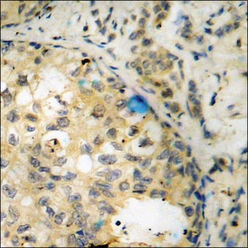
Anti-phospho-Histone H3 (Ser10) Antibody, Mitosis Marker is a Rabbit Polyclonal Antibody for detection of Histone H3 phosphorylated at serine 10.This highly published Ab, also known as Anti-H3S10p, has been validated in ICC, IP & WB.
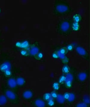
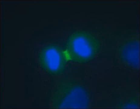
Immunocytochemistry Analysis: 1:500 dilution from a representative lot detected Histone H3 in HeLa and A431 cells.
Immunoprecipitation Analysis: 10 µg from a representative lot immunoprecipitated phospho Histone H3 (Ser10) from HeLa acid extract and Colcemid treated HeLa acid extracted RIPA lysate.
Beadlyte Assay Analysis: A representative lot recognizes Histone H3 phosphorylated on Ser10 by Luminex assay.
Research Category
Epigenetics & Nuclear Function
Research Sub Category
Histones

Quality

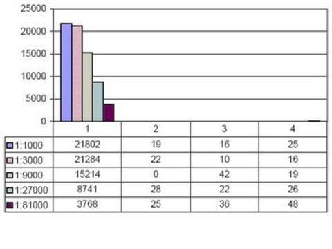
Evaluated by Western Blot in Colcemid treated HeLa acid extract.
Western Blot Analysis: 0.5 – 1 µg/ml of this antibody detected Histone H3 on 10 µg of Colcemid treated HeLa acid extract.

Target description

17 kDa

Physical form

Format: Purified
Protein A purified
Purified rabbit polyclonal serum in buffer containing 0.1 M Tris-glycine, pH 7.4, 0.150 mM NaCl and 0.05% sodium azide.

Storage and Stability

2-8°C for up to 6 months.

Analysis Note

Control
Colcemid treated HeLa acid extract

Legal Information

UPSTATE is a registered trademark of Merck KGaA, Darmstadt, Germany

Disclaimer

Unless otherwise stated in our catalog or other company documentation accompanying the product(s), our products are intended for research use only and are not to be used for any other purpose, which includes but is not limited to, unauthorized commercial uses, in vitro diagnostic uses, ex vivo or in vivo therapeutic uses or any type of consumption or application to humans or animals.

Not finding the right product?  

Try our Product Selector Tool.

Storage Class Code

10 - Combustible liquids

WGK

WGK 1


Certificates of Analysis (COA)

Search for Certificates of Analysis (COA) by entering the products Lot/Batch Number. Lot and Batch Numbers can be found on a product’s label following the words ‘Lot’ or ‘Batch’.

Already Own This Product?

Find documentation for the products that you have recently purchased in the Document Library.

Visit the Document Library

Context-specific requirements for Fgfr1 signaling through Frs2 and Frs3 during mouse development.
Hoch, RV; Soriano, P
Development null
Lyubomir T Vassilev et al.
Proceedings of the National Academy of Sciences of the United States of America, 103(28), 10660-10665 (2006-07-05)
CDK1 is a nonredundant cyclin-dependent kinase (CDK) with an essential role in mitosis, but its multiple functions still are poorly understood at a molecular level. Here we identify a selective small-molecule inhibitor of CDK1 that reversibly arrests human cells at
Overexpression of an N-terminally truncated isoform of the nuclear receptor coactivator amplified in breast cancer 1 leads to altered proliferation of mammary epithelial cells in transgenic mice.
Tilli, MT; Reiter, R; Oh, AS; Henke, RT; McDonnell, K; Gallicano, GI; Furth, PA; Riegel, AT
Molecular Endocrinology null
Loss of Hspa9b in zebrafish recapitulates the ineffective hematopoiesis of the myelodysplastic syndrome.
Craven, SE; French, D; Ye, W; de Sauvage, F; Rosenthal, A
Blood null
Identification and dynamics of two classes of aurora-like kinases in Arabidopsis and other plants.
Demidov, D; Van Damme, D; Geelen, D; Blattner, FR; Houben, A
Plant Cell null

Articles

Explore the basics of working with antibodies including technical information on structure, classes, and normal immunoglobulin ranges.

Explore the basics of working with antibodies including technical information on structure, classes, and normal immunoglobulin ranges.

Learn differences in monoclonal vs polyclonal antibodies including how antibodies are generated, clone numbers, and antibody formats.

Learn differences in monoclonal vs polyclonal antibodies including how antibodies are generated, clone numbers, and antibody formats.

See All

Our team of scientists has experience in all areas of research including Life Science, Material Science, Chemical Synthesis, Chromatography, Analytical and many others.

Contact Technical Service