Skip to Content
Merck
All Photos(2)

Documents

SAB4200721

Sigma-Aldrich

Anti-GABA antibody, Mouse monoclonal

clone GB-69, purified from hybridoma cell culture

Synonym(s):

Mouse Anti-Gamma-aminobutyric acid

Sign Into View Organizational & Contract Pricing


About This Item

UNSPSC Code:
12352203
NACRES:
NA.41

biological source

mouse

antibody form

purified from hybridoma cell culture

antibody product type

primary antibodies

clone

GB-69, monoclonal

form

buffered aqueous solution

species reactivity

rat, monkey, frog, mouse, human, gerbil

concentration

~1.0 mg/mL

technique(s)

ELISA: suitable
dot blot: suitable
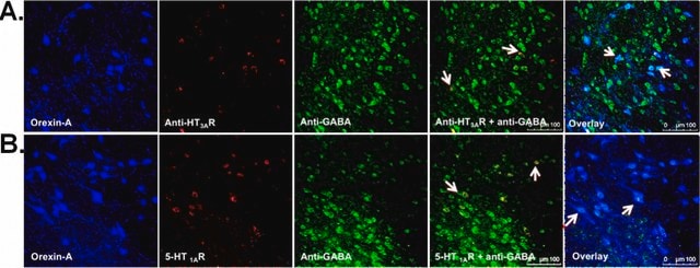
immunofluorescence: 5-10 μg/mL using human pancreatic tumor AsPC1 cell line
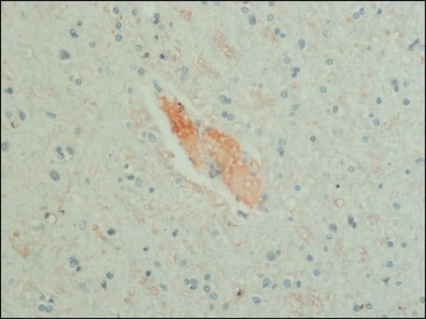
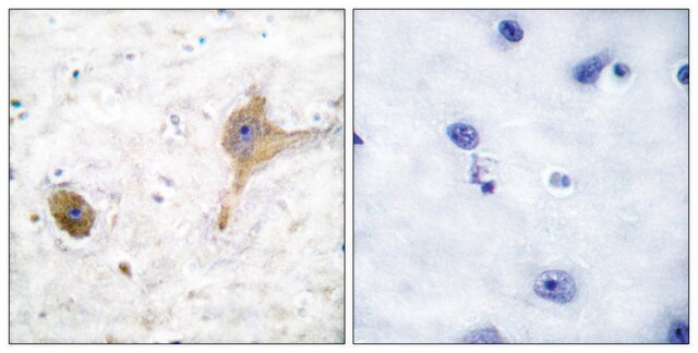
immunohistochemistry: 1-2.5 μg/mL using heat-retrieved formalin-fixed, paraffin-embedded human brain and/or cerebellum sections

isotype

IgG1

shipped in

dry ice

storage temp.

−20°C

target post-translational modification

unmodified

Gene Information

human ... GABRA1(2554)
mouse ... Gabra1(14394)
rat ... Gabra1(29705)
rhesus monkey ... Gabra1(574302)

General description

Anti-GABA antibody, Mouse monoclonal (mouse IgG1 isotype) is derived from the GB-69 hybridoma produced by the fusion of mouse myeloma cells and splenocytes from a mouse immunized with purified GABA conjugated to BSA. γ-Aminobutyric acid (GABA) is present in the central nervous system (CNS) of vertebrates. GABA is formed following enzymatic decarboxylation of L-glutamic acid by glutamic acid decarboxylase (GAD).

Immunogen

purified GABA conjugated to BSA

Application

Anti-GABA antibody, Mouse monoclonal has been used in:
  • immunofluorescence
  • immunohistochemistry
  • enzyme linked immunosorbent assay (ELISA)
  • dot-blot

Biochem/physiol Actions

γ-Aminobutyric acid (GABA) is the main inhibitory neurotransmitter. GABA mediates fast synaptic inhibition in the mature nervous system and plays multiple key roles as sensory circuits undergo functional development. GABA promotes proliferation of immune cells, embryonic stem cells and cortical progenitor cells. In addition, it is involved in protein synthesis and metabolism and plays a major role in the regulation of muscle tone, blood pressure, heart rate and respiration.

Physical form

Solution in 0.01 M phosphate buffered saline, pH 7.4, containing 15 mM sodium azide

Disclaimer

Unless otherwise stated in our catalog or other company documentation accompanying the product(s), our products are intended for research use only and are not to be used for any other purpose, which includes but is not limited to, unauthorized commercial uses, in vitro diagnostic uses, ex vivo or in vivo therapeutic uses or any type of consumption or application to humans or animals.

Not finding the right product?  

Try our Product Selector Tool.

Storage Class Code

10 - Combustible liquids

WGK

WGK 1

Flash Point(F)

Not applicable

Flash Point(C)

Not applicable


Certificates of Analysis (COA)

Search for Certificates of Analysis (COA) by entering the products Lot/Batch Number. Lot and Batch Numbers can be found on a product’s label following the words ‘Lot’ or ‘Batch’.

Already Own This Product?

Find documentation for the products that you have recently purchased in the Document Library.

Visit the Document Library

Distribution of glycine immunoreactivity in the brain of the Siberian sturgeon (Acipenser baeri): Comparison with gamma-aminobutyric acid
Adrio F, et al.
The Journal of Comparative Neurology, 519(6), 1115-1142 (2011)
GABA maintains the proliferation of progenitors in the developing chick ciliary marginal zone and non-pigmented ciliary epithelium
Ring H, et al.
PLoS ONE, 7(5), e36874-e36874 (2012)
Kunihiko Obata
Proceedings of the Japan Academy. Series B, Physical and biological sciences, 89(4), 139-156 (2013-04-12)
Signal transmission through synapses connecting two neurons is mediated by release of neurotransmitter from the presynaptic axon terminals and activation of its receptor at the postsynaptic neurons. γ-Aminobutyric acid (GABA), non-protein amino acid formed by decarboxylation of glutamic acid, is
Wei Fan et al.
eLife, 10 (2021-05-28)
Sphingolipids are important structural components of cell membranes and prominent signaling molecules controlling cell growth, differentiation, and apoptosis. Sphingolipids are particularly abundant in the brain, and defects in sphingolipid degradation are associated with several human neurodegenerative diseases. However, molecular mechanisms

Our team of scientists has experience in all areas of research including Life Science, Material Science, Chemical Synthesis, Chromatography, Analytical and many others.

Contact Technical Service