Skip to Content
Merck
All Photos(1)

Key Documents

316M-1

Sigma-Aldrich

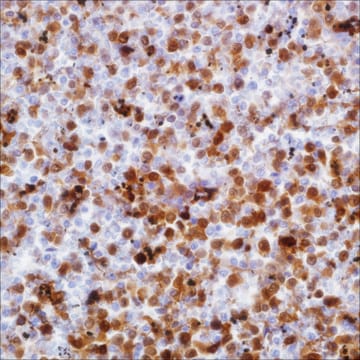
Perforin (MRQ-23) Mouse Monoclonal Antibody

Sign Into View Organizational & Contract Pricing


About This Item

UNSPSC Code:
12352203
NACRES:
NA.41

biological source

mouse

Quality Level

100
500

conjugate

unconjugated

antibody form

culture supernatant

antibody product type

primary antibodies

clone

MRQ-23, monoclonal

description

For In Vitro Diagnostic Use in Select Regions (See Chart)

form

buffered aqueous solution

species reactivity

human

packaging

vial of 0.1 mL concentrate (316M-14)
vial of 0.5 mL concentrate (316M-15)
bottle of 1.0 mL predilute (316M-17)
vial of 1.0 mL concentrate (316M-16)
bottle of 7.0 mL predilute (316M-18)

manufacturer/tradename

Cell Marque

technique(s)

immunohistochemistry (formalin-fixed, paraffin-embedded sections): 1:10-1:50

isotype

IgG1

control

spleen

shipped in

wet ice

storage temp.

2-8°C

Gene Information

human ... PRF1(5551)

General description

Perforin is a pore-forming protein that leads to osmotic lysis of the target cells and subsequently enables granzymes to enter the target cells and activate apoptosis, the cell death program. The expression of perforin is upregulated in activated CD8+ T-cells, but in NK cells the expression is constitutively very high and stable. Perforin expression can also be stimulated in some activated CD4+ T-cells. Although some investigators report a cytolytic potential of CD4+ T-cells, it appears more likely that CD8+ T-cells are the major effector population in Th1-associated inflammatory skin diseases. The role of perforin-mediated cytotoxicity has been demonstrated in various autoimmune diseases. In vitro and in vivo studies suggest that the cytotoxicity of CTLs may be mediated by cytotoxic granules in certain inflammatory diseases in humans. In addition, it seems that T-cell cytotoxicity against keratinocytes is mediated by perforin in some inflammatory skin diseases. Other authors suggest that perforin may have a dual role in alloimmune response (organ transplant applications). In one regard, it has a cytolytic function in acute rejection and, in contrast, it may be responsible for downregulating both CD4- and CD8-mediated alloimmune response.

Quality


IVD

IVD

IVD

RUO

Linkage

Perforin Positive Control Slides, Product No. 316S, are available for immunohistochemistry (formalin-fixed, paraffin-embedded sections).

Physical form

Solution in Tris Buffer, pH 7.3-7.7, with 1% BSA and <0.1% Sodium Azide

Preparation Note

Download the IFU specific to your product lot and formatNote: This requires a keycode which can be found on your packaging or product label.

Other Notes

For Technical Service please contact: 800-665-7284 or email: service@cellmarque.com

Legal Information

Cell Marque is a trademark of Merck KGaA, Darmstadt, Germany

Not finding the right product?  

Try our Product Selector Tool.


Certificates of Analysis (COA)

Search for Certificates of Analysis (COA) by entering the products Lot/Batch Number. Lot and Batch Numbers can be found on a product’s label following the words ‘Lot’ or ‘Batch’.

Already Own This Product?

Find documentation for the products that you have recently purchased in the Document Library.

Visit the Document Library

Iris Bittmann et al.
Virchows Archiv : an international journal of pathology, 445(4), 375-381 (2004-10-14)
Acute rejection and diffuse alveolar damage are major problems during the early time after transplantation. Against this background, lung biopsies after allogeneic lung transplantation were studied using immunohistochemistry. Biopsies with acute rejection, diffuse alveolar damage and morphological inconspicuous biopsies were
E S G d'Amore et al.
Pediatric and developmental pathology : the official journal of the Society for Pediatric Pathology and the Paediatric Pathology Society, 10(3), 181-191 (2007-05-31)
In this article, we describe the morphologic and immunophenotypic features of 75 cases of pediatric anaplastic large cell lymphoma (ALCL). According to the World Health Organization classification, 49 cases were common subtype ALCL, and respectively, 3, 6, and 17 cases
P G Chu et al.
Annals of diagnostic pathology, 3(2), 104-133 (1999-04-10)
Immunohistochemistry plays a key role in the diagnosis and classification of hematolymphoid neoplasms. New cell and lineage markers are constantly being discovered and added to the existing long list of antibodies. In this review article we provide general information and

Our team of scientists has experience in all areas of research including Life Science, Material Science, Chemical Synthesis, Chromatography, Analytical and many others.

Contact Technical Service