Saltar al contenido
Merck
Todas las fotos(2)

Documentos

MABN503

Sigma-Aldrich

Anti-WAVE-1 Antibody, clone K91/36

clone K91/36, from mouse

Sinónimos:

Wiskott-Aldrich syndrome protein family member 1, WASP family protein member 1, Protein WAVE-1, Verprolin homology domain-containing protein 1

Iniciar sesiónpara Ver la Fijación de precios por contrato y de la organización


About This Item

UNSPSC Code:
12352203
eCl@ss:
32160702
NACRES:
NA.41

biological source

mouse

Quality Level

antibody form

purified antibody

antibody product type

primary antibodies

clone

K91/36, monoclonal

species reactivity

human, mouse, rat

technique(s)

immunohistochemistry: suitable
western blot: suitable

isotype

IgG1κ

NCBI accession no.

UniProt accession no.

shipped in

wet ice

target post-translational modification

unmodified

Gene Information

human ... WASF1(8936)

General description

Protein WAVE-1 (WAVE1) is also known as Wiskott-Aldrich syndrome protein (WASP) family member 1 (WASF1), verprolin homology domain-containing protein 1, or SCAR1. WAVE1 is a downstream effector molecule involved in the transmission of signals from tyrosine kinase receptors and small GTPases to the actin cytoskeleton. WAVE1 binds actin and ARP2/3 complex. It binds the ARP2/3 complex through the C-terminal region and actin through verprolin homology (VPH) domain. WAVE1 is highly expressed in brain, with low expression in several other tissue.

Immunogen

Linear peptide corresponding to human WAVE-1.

Application

Detect WAVE1 using this mouse monoclonal antibody, Anti-WAVE-1 Antibody, clone K91/36 validated for use in western blotting & IHC.
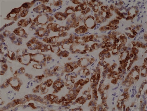
Immunohistochemistry Analysis: A representative lot detected WAVE-1 in adult rat hippocampal CA1 tissue (Prof. J. Trimmer, University of California, Davis).
Research Category
Neuroscience
Research Sub Category
Developmental Signaling

Quality

Evaluated by Western Blotting in rat brain tissue lysate.

Western Blotting Analysis: 1.0 µg/mL of this antibody detected WAVE-1 in 10 µg of rat brain tissue lysate.

Target description

~ 70 kDa observed

Physical form

Format: Purified
Protein G Purified
Purified mouse monoclonal IgG1κ in buffer containing 0.1 M Tris-Glycine (pH 7.4), 150 mM NaCl with 0.05% sodium azide.

Storage and Stability

Stable for 1 year at 2-8°C from date of receipt.

Analysis Note

Control
Rat brain tissue lysate

Other Notes

Concentration: Please refer to the Certificate of Analysis for the lot-specific concentration.

Disclaimer

Unless otherwise stated in our catalog or other company documentation accompanying the product(s), our products are intended for research use only and are not to be used for any other purpose, which includes but is not limited to, unauthorized commercial uses, in vitro diagnostic uses, ex vivo or in vivo therapeutic uses or any type of consumption or application to humans or animals.

¿No encuentra el producto adecuado?  

Pruebe nuestro Herramienta de selección de productos.

Storage Class

12 - Non Combustible Liquids

wgk_germany

WGK 1

flash_point_f

Not applicable

flash_point_c

Not applicable


Certificados de análisis (COA)

Busque Certificados de análisis (COA) introduciendo el número de lote del producto. Los números de lote se encuentran en la etiqueta del producto después de las palabras «Lot» o «Batch»

¿Ya tiene este producto?

Encuentre la documentación para los productos que ha comprado recientemente en la Biblioteca de documentos.

Visite la Librería de documentos

Jennifer Rakotomamonjy et al.
Cell reports, 42(8), 112845-112845 (2023-07-22)
Protocadherins (PCDHs) are cell adhesion molecules that regulate many essential neurodevelopmental processes related to neuronal maturation, dendritic arbor formation, axon pathfinding, and synaptic plasticity. Biallelic loss-of-function variants in PCDH12 are associated with several neurodevelopmental disorders (NDDs). Despite the highly deleterious outcome
Jessica M Hogestyn et al.
Molecular and cellular neurosciences, 123, 103770-103770 (2022-09-03)
Many neurodegenerative diseases have a multifactorial etiology and variable course of progression that cannot be explained by current models. Neurotropic viruses have long been suggested to play a role in these diseases, although their exact contributions remain unclear. Human herpesvirus
Binbin Li et al.
Development (Cambridge, England), 149(17) (2022-08-12)
Coordinated migration of the mesoderm is essential for accurate organization of the body plan during embryogenesis. However, little is known about how mesoderm migration influences posterior neural tube closure in mammals. Here, we show that spinal neural tube closure and
Wenqi Li et al.
The Journal of neuroscience : the official journal of the Society for Neuroscience, 36(44), 11283-11294 (2016-11-04)
Two distinct protein cofactors, p35 and p39, independently activate Cyclin-dependent kinase 5 (Cdk5), which plays diverse roles in normal brain function and the pathogenesis of many neurological diseases. The initial discovery that loss of p35 impairs neuronal migration in the

Nuestro equipo de científicos tiene experiencia en todas las áreas de investigación: Ciencias de la vida, Ciencia de los materiales, Síntesis química, Cromatografía, Analítica y muchas otras.

Póngase en contacto con el Servicio técnico