推荐产品
生物源
rabbit
品質等級
共軛
unconjugated
抗體表格
affinity isolated antibody
抗體產品種類
primary antibodies
無性繁殖
polyclonal
形狀
buffered aqueous solution
分子量
antigen ~75 kDa
物種活性
rat, human, mouse
包裝
antibody small pack of 25 μL
技術
immunoprecipitation (IP): 1-2 μL using human U87 cell lysate
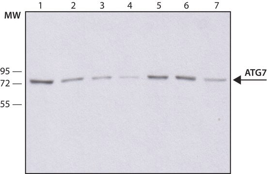
western blot: 0.5-1 μg/mL using whole extracts of mouse 3T3 cells
UniProt登錄號
運輸包裝
dry ice
儲存溫度
−20°C
目標翻譯後修改
unmodified
基因資訊
human ... ATG7(10533)
mouse ... Atg7(74244)
rat ... Atg7(312647)
一般說明
细胞自噬是细胞将部分细胞质送入溶酶体进行蛋白降解的代谢过程。自噬体形成中,ATG16基因至关重要,Atg7用于激活泛素样蛋白Atg12和Atg8。Atg7对于新生儿的氨基酸供应和饥饿诱导的小鼠蛋白质和细胞器的大量降解至关重要。ATG7抗体可用于免疫组化和免疫沉淀分析。兔抗ATG7抗体可与人、小鼠和大鼠的Atg7发生特异性反应。
免疫原
对应于人ATG7第38-50个氨基酸的合成肽,通过C-末端的半胱氨酸残基与KLH发生偶联。相应的序列在小鼠和大鼠中是相同的。
應用
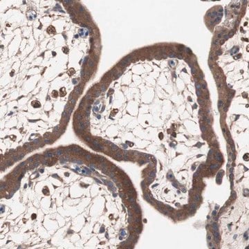
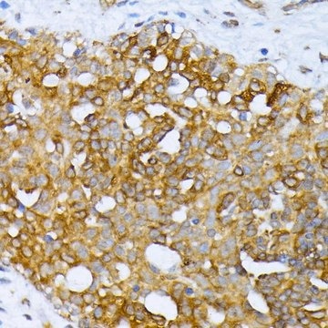
兔抗ATG7抗体适用于免疫印迹实验。还可用于人U87细胞裂解物的免疫沉淀实验(1-2μL)。
兔抗ATG7抗体适用:
- 自噬相关基因的实时定量聚合酶链反应
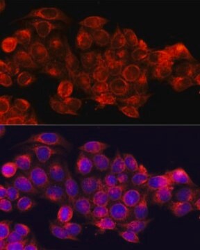
- 免疫荧光和免疫印迹实验
- 自噬蛋白的体内乙酰化/脱乙酰
在固定于4%多聚甲醛的小鼠中脑切片的免疫组化实验中,兔抗-ATG7抗体以1:2000稀释使用。
抗-ATG7抗体可以用于western blotting和免疫印迹。
生化/生理作用
自噬相关蛋白7(ATG7)调节caspase介导的自噬和细胞凋亡。这对肿瘤进展有显著作用,ATG7可充当肿瘤抑制剂。现已确知ATG7与许多类型的癌症有关,因此被认为是癌症治疗的靶标。ATG7基因的丢失有助于肝癌的发生。ATG7基因编码对于脂质化和偶联系统必不可少的E1样酶(E1-like enzyme):泛素样微管相关蛋白1A / 1B-轻链3(LC3)和自噬相关蛋白12(ATG12)。
外觀
0.01M 磷酸缓冲盐溶液,pH 7.4,含 15mM 叠氮化钠。
免責聲明
除非我们的产品目录或产品附带的其他公司文档另有说明,否则我们的产品仅供研究使用,不得用于任何其他目的,包括但不限于未经授权的商业用途、体外诊断用途、离体或体内治疗用途或任何类型的消费或应用于人类或动物。
未找到合适的产品?
试试我们的产品选型工具.
儲存類別代碼
12 - Non Combustible Liquids
水污染物質分類(WGK)
WGK 1
閃點(°F)
Not applicable
閃點(°C)
Not applicable
個人防護裝備
Eyeshields, Gloves, multi-purpose combination respirator cartridge (US)
其他客户在看
Midbody ring disposal by autophagy is a post-abscission event of cytokinesis
Pohl C and Jentsch S
Nature Cell Biology, 11(1), 65-65 (2009)
Man J Livingston et al.
Autophagy, 12(6), 976-998 (2016-04-29)
Renal fibrosis is the final, common pathway of end-stage renal disease. Whether and how autophagy contributes to renal fibrosis remains unclear. Here we first detected persistent autophagy in kidney proximal tubules in the renal fibrosis model of unilateral ureteral obstruction
The emerging links between sirtuins and autophagy
Sirtuins, 259-271 (2013)
Shinichi Kawano et al.
Biology open, 6(11), 1644-1653 (2017-10-04)
Adhesion of cells to the extracellular matrix (ECM) via focal adhesions (FAs) is crucial for cell survival, migration, and differentiation. Although the regulation of FAs, including by integrins and the ECM, is important to cell behavior, how FAs are regulated
Jessie Yanxiang Guo et al.
Genes & development, 27(13), 1447-1461 (2013-07-05)
Macroautophagy (autophagy hereafter) degrades and recycles proteins and organelles to support metabolism and survival in starvation. Oncogenic Ras up-regulates autophagy, and Ras-transformed cell lines require autophagy for mitochondrial function, stress survival, and engrafted tumor growth. Here, the essential autophagy gene
我们的科学家团队拥有各种研究领域经验,包括生命科学、材料科学、化学合成、色谱、分析及许多其他领域.
联系技术服务部门