
推荐产品


生物来源
mouse
质量水平
抗体形式
purified antibody
抗体产品类型
primary antibodies
克隆
XB6-AC5, monoclonal
分子量
calculated mol wt 41.6 kDa
observed mol wt ~32 kDa
纯化方式
using protein G
种属反应性
mouse, rat
包装
antibody small pack of 100
技术
immunocytochemistry: suitable
immunofluorescence: suitable
immunohistochemistry: suitable
immunoprecipitation (IP): suitable
western blot: suitable
同位素/亚型
IgG1κ
表位序列
N-terminal
Protein ID登记号
UniProt登记号
储存温度
2-8°C
基因信息
mouse ... Mlc1(170790)
特异性
Clone XB6-AC5 is a mouse monoclonal antibody that detects MLC1. It targets an epitope within 15 amino acids from the N-terminal region.
免疫原
A linear peptide corresponding to 15 amino acids from the N-terminal region of mouse MLC1.
应用
Quality Control Testing
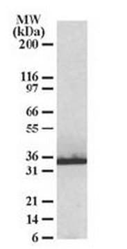
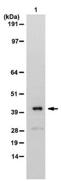
Evaluated by Western Blotting in wild-type Mouse brain membrane extract.
Western Blotting Analysis: A 1:1,000 dilution of this antibody detected MLC1 in Wild-type Mouse brain membrane extract, but not in extract from Mouse membrane with MLC1 knockout.
Tested Applications
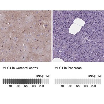
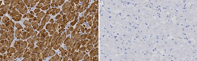
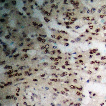
Immunohistochemistry: A representative lot detected MLC1 in Immunohistochemistry applications (Diaz-Castro, B., et al. (2019). Sci Transl Med. 11(514):eaaw8546; Gilbert, A., et al. (2019). Brain Struct Funct. 224(3):1267-1278; Sanchez, A., et al. (2020). Neurotherapeutics. 17(4):2041-2053).
Immunoprecipitation Analysis: Applications: A representative lot detected MLC1 in Immunohistochemistry applications (Elorza-Vidal, X., et al. (2018). Neurobiol Dis. 119:88-99).
Immunofluorescence Analysis (IF): A representative lot detected MLC1 in Immunofluorescence applications (Sirisi, S., et al. (2017). Hum Mol Genet.;26(13) 2436-2450; Elorza-Vidal, X., et al. (2018). Neurobiol Dis. 119:88-99; Perez-Rius, C., et al. (2019). Orphanet J Rare Dis. 14(1):268; Sanchez, A., et al. (2020). Neurotherapeutics. 17(4):2041-2053).
Immunocytochemistry Analysis: A representative lot detected MLC1 in Immunocytochemistry applications (Sirisi, S., et al. (2017). Hum Mol Genet. 26(13):2436-2450; Elorza-Vidal, X., et al. (2018). Neurobiol Dis. 119:88-99).
Western Blotting Analysis: A representative lot detected MLC1 in Western Blotting applications (Sirisi, S., et al. (2017). Hum Mol Genet. 26(13):2436-2450; Elorza-Vidal, X., et al. (2018). Neurobiol Dis. 119:88-99; Gilbert, A., et al. (2019). Brain Struct Funct. 224(3):1267-1278; Perez-Rius, C., et al. (2019). Orphanet J Rare Dis. 14(1):268; Sanchez, A., et al. (2020). Neurotherapeutics. 17(4):2041-2053).
Note: Actual optimal working dilutions must be determined by end user as specimens, and experimental conditions may vary with the end user.
Evaluated by Western Blotting in wild-type Mouse brain membrane extract.
Western Blotting Analysis: A 1:1,000 dilution of this antibody detected MLC1 in Wild-type Mouse brain membrane extract, but not in extract from Mouse membrane with MLC1 knockout.
Tested Applications
Immunohistochemistry: A representative lot detected MLC1 in Immunohistochemistry applications (Diaz-Castro, B., et al. (2019). Sci Transl Med. 11(514):eaaw8546; Gilbert, A., et al. (2019). Brain Struct Funct. 224(3):1267-1278; Sanchez, A., et al. (2020). Neurotherapeutics. 17(4):2041-2053).
Immunoprecipitation Analysis: Applications: A representative lot detected MLC1 in Immunohistochemistry applications (Elorza-Vidal, X., et al. (2018). Neurobiol Dis. 119:88-99).
Immunofluorescence Analysis (IF): A representative lot detected MLC1 in Immunofluorescence applications (Sirisi, S., et al. (2017). Hum Mol Genet.;26(13) 2436-2450; Elorza-Vidal, X., et al. (2018). Neurobiol Dis. 119:88-99; Perez-Rius, C., et al. (2019). Orphanet J Rare Dis. 14(1):268; Sanchez, A., et al. (2020). Neurotherapeutics. 17(4):2041-2053).
Immunocytochemistry Analysis: A representative lot detected MLC1 in Immunocytochemistry applications (Sirisi, S., et al. (2017). Hum Mol Genet. 26(13):2436-2450; Elorza-Vidal, X., et al. (2018). Neurobiol Dis. 119:88-99).
Western Blotting Analysis: A representative lot detected MLC1 in Western Blotting applications (Sirisi, S., et al. (2017). Hum Mol Genet. 26(13):2436-2450; Elorza-Vidal, X., et al. (2018). Neurobiol Dis. 119:88-99; Gilbert, A., et al. (2019). Brain Struct Funct. 224(3):1267-1278; Perez-Rius, C., et al. (2019). Orphanet J Rare Dis. 14(1):268; Sanchez, A., et al. (2020). Neurotherapeutics. 17(4):2041-2053).
Note: Actual optimal working dilutions must be determined by end user as specimens, and experimental conditions may vary with the end user.
目标描述
Membrane protein MLC1 (UniProt: Q8VHK5; also known as MLC1) is encoded by the Mlc1 gene (Gene ID: 170790) in murine species. MLC1 is a multi-pass membrane protein that regulates the response of astrocytes to hypo-osmosis by promoting calcium influx. It is a highly hydrophobic protein with eight transmembrane domains and short amino and carboxylic- cytoplasmic tails. It forms highly stable dimeric and oligomeric structures. Within the brain, it is mainly expressed in astrocytes, particularly at the astrocyte end-feet contacting the blood-brain barrier and the pial membrane. MLC1 may also be involved in transporting molecules across the blood-brain barrier and the brain-cerebrospinal fluid barrier. Its higher expression in astrocytes contacting blood vessels suggest its role in the regulation of ion and water homeostasis. Studies have shown that MLC1 establishes structural and/or functional interactions with several ion/water channels and transporters and ion channel accessory proteins. These interactions are affected by mutations in Mlc1 gene that cause Megalencephalic leukoencephalopathy with subcortical cysts (MLC) that is characterized mainly by myelin vacuolization and early onset of macrocephaly, early in life. GlialCAM is reported to be essential for MLC1 endoplasmic reticulum exit and targeting to astrocyte-astrocyte junctions. Mutations in Glial CAM are also observed in about 50% of subjects with MLC. MLC1 is phosphorylated at its N- and C-terminal regions by both PKA and PKC and PKC alone, respectively. Treatment with agents that activate PKC or PKA or with phosphatase inhibitors is reported to modify MLC1 plasma membrane expression and formation of multimeric structures. (Ref.: Elorza-Vidal, X., et al. (2018). Neurobiol. Dis. 119; 88-99; Sirisi, S., et al. (2017). Hum. Mol. Genet. 26(13); 2436-2450; Brignone, MS., et al. (2015). Front. Cell. Neurosci. 9; 66).
外形
Purified mouse monoclonal antibody IgG1 in buffer containing 0.1 M Tris-Glycine (pH 7.4), 150 mM NaCl with 0.05% sodium azide.
重悬
1.0 mg/mL. Please refer to guidance on suggested starting dilutions and/or titers per application and sample type.
储存及稳定性
Recommended storage: +2°C to +8°C.
其他说明
Concentration: Please refer to the Certificate of Analysis for the lot-specific concentration.
免责声明
Unless otherwise stated in our catalog or other company documentation accompanying the product(s), our products are intended for research use only and are not to be used for any other purpose, which includes but is not limited to, unauthorized commercial uses, in vitro diagnostic uses, ex vivo or in vivo therapeutic uses or any type of consumption or application to humans or animals.
未找到合适的产品?
试试我们的产品选型工具.
储存分类代码
12 - Non Combustible Liquids
WGK
WGK 1
闪点(°F)
Not applicable
闪点(°C)
Not applicable
我们的科学家团队拥有各种研究领域经验,包括生命科学、材料科学、化学合成、色谱、分析及许多其他领域.
联系技术服务部门