Accéder au contenu
Merck
Toutes les photos(3)

Principaux documents

09-403

Sigma-Aldrich

Anti-TKS5 (SH3 #1) Antibody

from rabbit

Synonyme(s) :

Adaptor protein TKS5, Five SH3 domain-containing protein, SH3 and PX domains 2A, SH3 multiple domains 1, SH3 multiple domains protein 1, five SH3 domains

Se connecterpour consulter vos tarifs contractuels et ceux de votre entreprise/organisme


About This Item

Code UNSPSC :
12352203
eCl@ss :
32160702
Nomenclature NACRES :
NA.41

Source biologique

rabbit

Niveau de qualité

Forme d'anticorps

purified antibody

Type de produit anticorps

primary antibodies

Clone

polyclonal

Espèces réactives

mouse, human

Technique(s)

immunocytochemistry: suitable
immunoprecipitation (IP): suitable
western blot: suitable

Numéro d'accès NCBI

Numéro d'accès UniProt

Conditions d'expédition

wet ice

Modification post-traductionnelle de la cible

unmodified

Informations sur le gène

Description générale

Tks5 (previously known as Fish), is a large scaffolding protein with one amino-terminal PX domain and five SH3 domains. It is cytoplasmic in normal fibroblasts. In Src-transformed cells, Tks5 localizes to podosomes. The coexpression of Tks5 and constitutively active Src in epithelial cancer cells resulted in the appearance of podosomes/invadopodia. Cells with reduced Tks5 expression are poorly invasive through matrigel. Tks5 is expressed and localized to podosomes/invadopodia in some invasive human cancer cell lines and tumor tissue, particularly breast cancers and melanomas. Thus, Tks5 appears to be required for podosome/invadopodia formation and for invasion of some cancer cells.

Spécificité

Other homologies: Human, Canine, Equine, Rat, and Bovine (100% sequence homology), Chicken (93% sequence homology)

Immunogène

Epitope: 166-225 aa
Recombinant Protein, aa: 166-225. Homology: Human, Dog, Horse, Rat, Cow (100%, 60/60); Chicken (93%, 56/60)

Application

Anti-TKS5 (SH3 #1) Antibody detects level of TKS5 (SH3 #1) & has been published & validated for use in WB, IC & IP.
Research Category
Cell Structure
Research Sub Category
Cytoskeletal Signaling
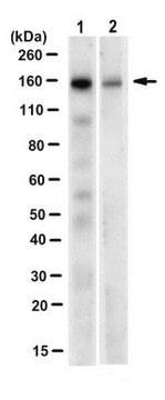
Western Blot Analysis: Antibody detected TKS5 in 20 μg of NIH-3T3 (100% confluent) lysate.

Qualité

Routinely evaluated by Western Blot on NIH-3T3 (100% confluent) lysate.

Description de la cible

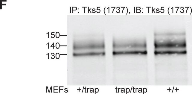
130-150 kDa Uncharacterized bands(s) may be observed in some cell lysates.

Forme physique

Format: Purified
Protein A purified
Purified in 0.1M Tris-Glycine (pH7.4) 150mM NaCl with 0.05% NaN3.

Stockage et stabilité

Maintain at 2-8°C in undiluted aliquots for up to 1 year after date of receipt.

Remarque sur l'analyse

Control
Western Blot::
NIH-3T3 (100% confluent) lysate
Immunocytochemistry:
Mouse 3T3-Src(Y527F) cells

Autres remarques

Concentration: Please refer to the Certificate of Analysis for the lot-specific concentration.

Clause de non-responsabilité

Unless otherwise stated in our catalog or other company documentation accompanying the product(s), our products are intended for research use only and are not to be used for any other purpose, which includes but is not limited to, unauthorized commercial uses, in vitro diagnostic uses, ex vivo or in vivo therapeutic uses or any type of consumption or application to humans or animals.

Vous ne trouvez pas le bon produit ?  

Essayez notre Outil de sélection de produits.

Code de la classe de stockage

12 - Non Combustible Liquids

Classe de danger pour l'eau (WGK)

WGK 1

Point d'éclair (°F)

Not applicable

Point d'éclair (°C)

Not applicable


Certificats d'analyse (COA)

Recherchez un Certificats d'analyse (COA) en saisissant le numéro de lot du produit. Les numéros de lot figurent sur l'étiquette du produit après les mots "Lot" ou "Batch".

Déjà en possession de ce produit ?

Retrouvez la documentation relative aux produits que vous avez récemment achetés dans la Bibliothèque de documents.

Consulter la Bibliothèque de documents

Karla C Williams et al.
Oncogene, 38(19), 3598-3615 (2019-01-18)
Invadopodia are cell protrusions that mediate cancer cell extravasation but the microenvironmental cues and signaling factors that induce invadopodia formation during extravasation remain unclear. Using intravital imaging and loss of function experiments, we determined invadopodia contain receptors involved in chemotaxis
Samuel J Ghilardi et al.
Molecular biology of the cell, 32(18), 1707-1723 (2021-07-01)
Interactions between the actin cytoskeleton and the plasma membrane are important in many eukaryotic cellular processes. During these processes, actin structures deform the cell membrane outward by applying forces parallel to the fiber's major axis (as in migration) or they
Nawal Bendris et al.
Journal of cell science, 129(14), 2804-2816 (2016-06-10)
The ability of cancer cells to degrade the extracellular matrix and invade interstitial tissues contributes to their metastatic potential. We recently showed that overexpression of sorting nexin 9 (SNX9) leads to increased cell invasion and metastasis in animal models, which
Deciphering the involvement of the Hippo pathway co-regulators, YAP/TAZ in invadopodia formation and matrix degradation.
Venghateri, et al.
Cell Death & Disease, 14, 290-290 (2023)
Sara K Donnelly et al.
The Journal of cell biology, 216(12), 4331-4349 (2017-10-25)
The initial step of metastasis is the local invasion of tumor cells into the surrounding tissue. Invadopodia are actin-based protrusions that mediate the matrix degradation necessary for invasion and metastasis of tumor cells. We demonstrate that Rac3 GTPase is critical

Notre équipe de scientifiques dispose d'une expérience dans tous les secteurs de la recherche, notamment en sciences de la vie, science des matériaux, synthèse chimique, chromatographie, analyse et dans de nombreux autres domaines..

Contacter notre Service technique