Direkt zum Inhalt
Merck

281M-8

Sigma-Aldrich

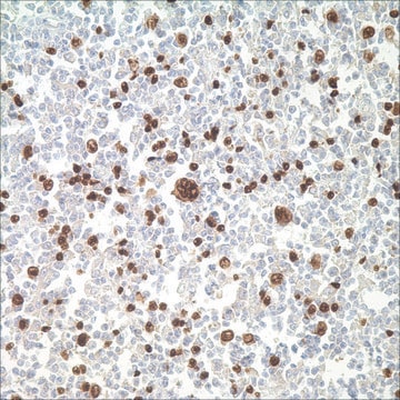
MART-1 (Melan A) (A103) Mouse Monoclonal Antibody

Anmeldenzur Ansicht organisationsspezifischer und vertraglich vereinbarter Preise


About This Item

UNSPSC-Code:
12352203
NACRES:
NA.41

Biologische Quelle

mouse

Qualitätsniveau

100
500

Konjugat

unconjugated

Antikörperform

culture supernatant

Antikörper-Produkttyp

primary antibodies

Klon

A103, monoclonal

Beschreibung

For In Vitro Diagnostic Use in Select Regions (See Chart)

Form

buffered aqueous solution

Speziesreaktivität

human

Verpackung

vial of 0.1 mL concentrate (281M-84)
vial of 0.5 mL concentrate (281M-85)
bottle of 1.0 mL predilute (281M-87)
vial of 1.0 mL concentrate (281M-86)
bottle of 7.0 mL predilute (281M-88)

Hersteller/Markenname

Cell Marque

Methode(n)

immunohistochemistry (formalin-fixed, paraffin-embedded sections): 1:100-1:500

Isotyp

IgG1

Kontrolle

melanoma

Versandbedingung

wet ice

Lagertemp.

2-8°C

Visualisierung

cytoplasmic

Angaben zum Gen

human ... MLANA(2315)

Allgemeine Beschreibung

MART-1 (auch als Melan A bekannt) ist ein Melanom-Antigen, das von T-Zellen erkannt wird.1-2 MART-1 ist ein Transmembranprotein, das in Melanozyten von normaler Haut, Nävi und den meisten Melanomen vorkommt. MART-1 ist ein nützlicher Marker zum Identifizieren von Melanomen.

Qualität


IVD

IVD

IVD

RUO

Verlinkung

MART-1-positive Kontrollobjektträger, Produktnr. 281S, sind für den Einsatz in der Immunhistochemie (formalinfixierte Paraffinschnitte) erhältlich.

Physikalische Form

Lösung in Tris-Puffer, pH 7,3-7,7, mit 1 % BSA und <0,1 % Natriumazid

Angaben zur Herstellung

Laden Sie die für Ihre Produktcharge und Ihr Format geltende Gebrauchsanweisung herunterHinweis: Dies erfordert einen Schlüsselcode, der auf Ihrer Verpackung oder Ihrem Produktetikett angegeben ist.

Sonstige Hinweise

Den Technischen Kundendienst erreichen Sie unter: 800-665-7284 oder per E-Mail: service@cellmarque.com

Rechtliche Hinweise

Cell Marque is a trademark of Merck KGaA, Darmstadt, Germany

Sie haben nicht das passende Produkt gefunden?  

Probieren Sie unser Produkt-Auswahlhilfe. aus.


Analysenzertifikate (COA)

Suchen Sie nach Analysenzertifikate (COA), indem Sie die Lot-/Chargennummer des Produkts eingeben. Lot- und Chargennummern sind auf dem Produktetikett hinter den Wörtern ‘Lot’ oder ‘Batch’ (Lot oder Charge) zu finden.

Besitzen Sie dieses Produkt bereits?

In der Dokumentenbibliothek finden Sie die Dokumentation zu den Produkten, die Sie kürzlich erworben haben.

Die Dokumentenbibliothek aufrufen

Cynthia Kucher et al.
The American Journal of dermatopathology, 26(6), 452-457 (2004-12-25)
Desmoplastic melanoma (DMM) is an uncommon melanoma variant with a distinct morphology, including a prominent spindle cell component with fibrosis, as well as a distinct immunohistochemical profile. Histologically, the spindle cell component of DMM can be confused with sclerotic/desmoplastic nevi
Ravi Suchak et al.
The American Journal of dermatopathology, 36(5), 387-391 (2014-01-08)
To assess the usefulness of routine melan-A immunohistochemistry (IHC) for exclusion of microinvasion in lentigo maligna (LM). One hundred and twenty cases of LM from our archives were reviewed by 2 authors with S100 protein and melan-A IHC using a
P G Coulie et al.
The Journal of experimental medicine, 180(1), 35-42 (1994-07-01)
It has been reported previously that antitumor cytolytic T lymphocyte (CTL) clones can be isolated from blood lymphocytes of HLA-A2 melanoma patients, after stimulation in vitro with autologous tumor cells, and that some of these CTL clones lyse most HLA-A2
Y Kawakami et al.
Proceedings of the National Academy of Sciences of the United States of America, 91(9), 3515-3519 (1994-04-26)
By cDNA expression cloning we have isolated a gene encoding a shared human melanoma antigen recognized by HLA-A2 restricted autologous and allogenic tumor-infiltrating lymphocytes (TILs) from patients with metastatic melanoma. By using both transient and stable expression systems, transfection of
Klaus Helm et al.
Journal of cutaneous pathology, 35(10), 931-934 (2008-05-23)
Distinguishing lentigo maligna from solar lentigo, and pigmented actinic keratosis can sometimes be problematic. Melan-A is an immunohistochemical marker which that can be helpful in decorating the melanocytes of pigmented lesions. A recent report has suggested that Melan-A may spuriously

Unser Team von Wissenschaftlern verfügt über Erfahrung in allen Forschungsbereichen einschließlich Life Science, Materialwissenschaften, chemischer Synthese, Chromatographie, Analytik und vielen mehr..

Setzen Sie sich mit dem technischen Dienst in Verbindung.