Skip to Content
Merck
All Photos(6)

Key Documents

AB175

Sigma-Aldrich

Anti-GABA Antibody

serum, Chemicon®

Synonym(s):

Anti-GABA Receptor Antibody, GABA Receptor Antibody

Sign Into View Organizational & Contract Pricing


About This Item

UNSPSC Code:
12352203
eCl@ss:
32160702
NACRES:
NA.41

biological source

guinea pig

Quality Level

antibody form

serum

antibody product type

primary antibodies

clone

polyclonal

species reactivity

rat

species reactivity (predicted by homology)

all

manufacturer/tradename

Chemicon®

technique(s)

ELISA: suitable
immunohistochemistry: suitable (paraffin)

shipped in

dry ice

target post-translational modification

unmodified

Gene Information

General description

GABA (gamma-aminobutyric acid) is the major inhibitory neurotransmitter in the central nervous system that inhibits the generation of the action potential of the neuron. It is involved in the pathogenesis of certain neurological and psychiatric disorders. GABA is produced from glutamic acid in a reaction catalyzed by glutamic acid decarboxylase. In this reaction pyridoxal phosphate serves as a co-factor. GABA interacts with the GABAA and GABAB receptors, which are widely distributed throughout the nervous system and in a variety of cell types. They differ in their pharmacological, electrophysiological, and biochemical properties. GABAA-receptor complex mediates an increase in membrane conductance with an equilibrium potential near the resting level of −70 mV. This conductance increase often is accompanied by a membrane hyperpolarization, which later results in a reduction in the probability of action potential initiation. The reduction in membrane resistance is accomplished by the GABA-dependent facilitation of Cl− ion influx. GABAB receptors are coupled indirectly to K+ channels. When activated, GABAB receptors reduce Calcium ion conductance and inhibit cAMP production via intracellular mechanisms mediated by G-proteins. GABAB receptors are known to mediate both postsynaptic and presynaptic inhibition.

Specificity

GABA is a neurotransmitter synthesized in all species, and is therefore expected to react to all species.
Recognizes GABA. Staining was blocked by preabsorbing with 100 μM GABA conjugated to glutaraldehyde. 500 μM of similar conjugations of glutamic acid, glutamate and taurine failed to block staining.

Immunogen

BSA-conjugated GABA-Glutaraldehyde

Application

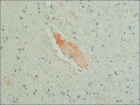
Immunohistochemistry Analysis: A 1:500 dilution from a representative lot detected GABA in rat brain tissue.

ELISA Analysis: A representative lot from an independent laboratory detected GABA in a competitive ELISA.

Immunohistochemistry Protocol:

Tissues fixed with 4% paraformaldehyde and 0-0.5% glutaraldehyde gives good results. Glutaraldehyde is required for antibody reactivity.

1) Tissue is fixed with 4% paraformaldehyde, 0-0.5% glutaraldehyde, 0.5% potassium dichromate in 0.1M phosphate buffer at pH 6.5.

2) Tissue is post-fixed overnight, vibratome sectioned in 50 mm and incubated in 0.05M Tris buffer, pH 6.5 for three hours.

3) Sections are incubated for 18-24 hours in AB175 diluted in PBS containing 0.1% sodium azide, 0.2% Triton X-100 and 1% normal goat serum.

4) Fluorescein conjugated antibody or ABC system may be used as the secondary reagent.

Note: Without colchicine pretreatment well-stained cell bodies are visible in the cerebral cortex, cerebrallar cortex, superior colliculus and some brainstem raphe. With colchicine pretreatment, additional cell body staining is present in the interpeduncular nucleus and the dorsal column nuclei.
Research Category
Neuroscience
Research Sub Category
Neurotransmitters & Receptors
This Anti-GABA Antibody is validated for use in Immunohistochemistry (Paraffin) and Enzyme Immunoassay (ELISA) for the detection of GABA.

Quality

Evaluated by Immunohistochemistry in rat brain tissue.

Immunohistochemistry Analysis: A 1:500 dilution of this antibody detected GABA in rat brain tissue.

Physical form

Depleted serum
Guinea Pig polyclonal BSA-depleted antiserum with 0.05% sodium azide.

Storage and Stability

Stable for 1 year at -20°C from date of receipt.
Handling Recommendations: Upon receipt and prior to removing the cap, centrifuge the vial and gently mix the solution. Aliquot into microcentrifuge tubes and store at -20°C. Avoid repeated freeze/thaw cycles, which may damage IgG and affect product performance.

Analysis Note

Control
Rat Brain tissue

Other Notes

Concentration: Variable

Legal Information

CHEMICON is a registered trademark of Merck KGaA, Darmstadt, Germany

Disclaimer

Unless otherwise stated in our catalog or other company documentation accompanying the product(s), our products are intended for research use only and are not to be used for any other purpose, which includes but is not limited to, unauthorized commercial uses, in vitro diagnostic uses, ex vivo or in vivo therapeutic uses or any type of consumption or application to humans or animals.

Not finding the right product?  

Try our Product Selector Tool.

Storage Class Code

12 - Non Combustible Liquids

WGK

WGK 1

Flash Point(F)

Not applicable

Flash Point(C)

Not applicable


Certificates of Analysis (COA)

Search for Certificates of Analysis (COA) by entering the products Lot/Batch Number. Lot and Batch Numbers can be found on a product’s label following the words ‘Lot’ or ‘Batch’.

Already Own This Product?

Find documentation for the products that you have recently purchased in the Document Library.

Visit the Document Library

Ji-Jie Pang et al.
Investigative ophthalmology & visual science, 52(7), 4886-4896 (2011-04-13)
To examine the specificity and reliability of a retrograde double-labeling technique that was recently established for identification of retinal ganglion cells (GCs) and to characterize the morphology of displaced (d)GCs (dGs). A mixture of the gap-junction-impermeable dye Lucifer yellow (LY)
Kristina D Micheva et al.
eNeuro, 5(5) (2018-11-09)
Numerous types of inhibitory neurons sculpt the performance of human neocortical circuits, with each type exhibiting a constellation of subcellular phenotypic features in support of its specialized functions. Axonal myelination has been absent among the characteristics used to distinguish inhibitory
NaHye Lee et al.
Experimental neurobiology, 27(5), 365-376 (2018-11-16)
Medium-chain fatty acids (MCFAs) are mostly generated from dietary triglycerides and can penetrate the blood-brain barrier. Astrocytes in the brain use MCFAs as an alternative energy source. In addition, MCFAs have various regulatory and signaling functions in astrocytes. However, it
Anatomical characterization of a rabbit cerebellar eyeblink premotor pathway using pseudorabies and identification of a local modulatory network in anterior interpositus.
Gonzalez-Joekes, J; Schreurs, BG
The Journal of Neuroscience null
Mei Chen et al.
PloS one, 8(4), e61381-e61381 (2013-05-03)
Previous studies have shown that CCL2/CX3CR1 deficient mice on C57BL/6N background (with rd8 mutation) have an early onset (6 weeks) of spontaneous retinal degeneration. In this study, we generated CCL2(-/-)CX3CR1(GFP/GFP) mice on the C57BL/6J background. Retinal degeneration was not detected

Our team of scientists has experience in all areas of research including Life Science, Material Science, Chemical Synthesis, Chromatography, Analytical and many others.

Contact Technical Service