
제품 SAB5500134은(는)귀하의 국가에서 현재 판매되지 않습니다. 고객지원팀으로 연락바랍니다.
추천 제품


생물학적 소스
rabbit
Quality Level
재조합
expressed in proprietary host
결합
unconjugated
항체 형태
tissue culture supernatant
항체 생산 유형
primary antibodies
클론
SP6, monoclonal
종 반응성
human (tested)
종 반응성(상동성에 의해 예측)
bovine
기술
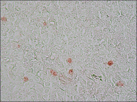
immunohistochemistry: 1:200
동형
IgG
UniProt 수납 번호
배송 상태
wet ice
저장 온도
2-8°C
타겟 번역 후 변형
unmodified
유전자 정보
human ... MKI67(4288)
일반 설명
Ki-67 is a nuclear protein, which is expressed in the proliferating cells. Ki-67 is preferentially expressed during late G1-, S-, M-, and G2-phases of the cell cycle, while cells in the G0 (quiescent) phase are negative for this protein.
The gene marker of proliferation Ki-67 (MKI67), spanning 15 exons, is mapped to human chromosome 10q26.2. The encoded protein contains phosphopeptide-binding forkhead-associated (FHA) domain and a protein phosphatase 1 (PP1)-binding site at N- terminal. It possesses 16 tandem repeats at central region and leucine and arginine (LR) pairs at C- terminal end.
면역원
Synthetic peptide from C-terminus of human Ki-67 protein.
애플리케이션
Anti-KI-67 antibody, Rabbit monoclonal has been used in immunohistochemistry.
생화학적/생리학적 작용
Marker of proliferation Ki-67 (MKI67), which is a part of the mitotic chromosome periphery, functions as a biological surfactant to maintain individual mitotic chromosomes dispersed in the cytoplasm after nuclear envelope disassembly. Ki-67 is a vital prognostic marker in breast and prostate cancer. Expression of the encoded protein is associated with cell-proliferation rate. Ki-67 is expressed at all active phases of the cell cycle (G (1), S, G (2), and mitosis), except resting cells (G (0)). Therefore, this protein is considered to be a potent cell proliferation marker.
특징 및 장점
Evaluate our antibodies with complete peace of mind. If the antibody does not perform in your application, we will issue a full credit or replacement antibody. Learn more.
물리적 형태
0.1 ml rabbit monoclonal antibody supplied as tissue culture supernatant in TBS/1% BSA buffer pH 7.5 with less than 0.1% sodium azide.
면책조항
Unless otherwise stated in our catalog or other company documentation accompanying the product(s), our products are intended for research use only and are not to be used for any other purpose, which includes but is not limited to, unauthorized commercial uses, in vitro diagnostic uses, ex vivo or in vivo therapeutic uses or any type of consumption or application to humans or animals.
적합한 제품을 찾을 수 없으신가요?
당사의 제품 선택기 도구.을(를) 시도해 보세요.
Storage Class Code
10 - Combustible liquids
WGK
WGK 2
Flash Point (°F)
Not applicable
Flash Point (°C)
Not applicable
가장 최신 버전 중 하나를 선택하세요:
시험 성적서(COA)
Lot/Batch Number
Tal Falick Michaeli et al.
Proceedings of the National Academy of Sciences of the United States of America, 119(52), e2212306119-e2212306119 (2022-12-20)
Injury to muscle brings about the activation of stem cells, which then generate new myocytes to replace damaged tissue. We demonstrate that this activation is accompanied by a dramatic change in the stem-cell methylation pattern that prepares them epigenetically for
Job Komen et al.
Micromachines, 13(5) (2022-05-29)
The cancer xenograft model in which human cancer cells are implanted in a mouse is one of the most used preclinical models to test the efficacy of novel cancer drugs. However, the model is imperfect; animal models are ethically burdened
Janneke F Linnekamp et al.
Cancers, 13(10) (2021-06-03)
DNA hypermethylation is common in colon cancer. Previously, we have shown that methylation of WNT target genes predicts poor prognosis in stage II colon cancer. The primary objective of this study was to assess whether pre-operative treatment with decitabine can
Madhavi Joshi et al.
Behavioural brain research, 372, 112029-112029 (2019-06-14)
Hypoglycemia induced brain injury poses a major setback to optimal blood glucose regulation during diabetes. It causes irreversible injury in several brain regions culminating in improper function. Neuregulin 1 and ErbB receptors are involved in regeneration during adulthood as well
The familial dysautonomia disease gene IKBKAP is required in the developing and adult mouse central nervous system.
Chaverra M, et al.
Disease models & mechanisms, 10(5), 605-618 (2017)
활성 필터
자사의 과학자팀은 생명 과학, 재료 과학, 화학 합성, 크로마토그래피, 분석 및 기타 많은 영역을 포함한 모든 과학 분야에 경험이 있습니다..
고객지원팀으로 연락바랍니다.