
제품 MABT456은(는)귀하의 국가에서 현재 판매되지 않습니다. 고객지원팀으로 연락바랍니다.
추천 제품


일반 설명
Moesin (membrane-organizing extension spike protein) is the protein encoded by the MSN gene. The MSN encoded protein moesin is a member of the ERM (ezrin, moesin, radixin) family of proteins that regulate linkage between the plasma membrane and the actin cytoskeleton. Thus, the MSN encoded protein is a peripheral membrane protein that is keenly involved in the signaling and structural connections in cytoskeletal plasma membrane structures like microvilli. Moesin in its active, phosphorylated state strongly binds actin and helps anchor it to the plasma membrane. The MSN encoded Moesin is localized to filopodia and other membranous extensions of various cell types and thus plays a role in cell-cell recognition signaling, cell movement, and cell transport. In its inactive, non-phosphorylated state, Moesin binds itself in a head-tail fashion and is incapable of binding actin or other cytoskeletal proteins. The kinase ROCK2 is central to the opening up of moesin and allowing its interaction with actin and the plasma membrane. EMD-Millipore’s Anti-MSN, clone 2C12 has been tested in western blots against purified recombinant protein as well as multiple human cancer cell lysates. The clone has also been tested successfully in paraffin embedded immunohistochemistry on human colon tissue and by flow cytometry on human Jurkat cells and ELISA using purified recombinant antigen.
면역원
Purified protein fragment of human MSN expressed in E. coli.
애플리케이션
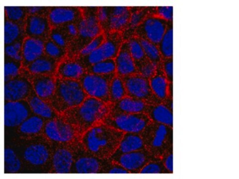
Detect Moesin using this mouse monoclonal antibody, Anti-MSN Antibody, clone 2C12 validated for use in western blotting, IHC & Flow Cytometry.
Immunohistochemistry Analysis: A 1:100-1,000 dilution from a representative lot detected MSN in human tonsil and colon tissues.
Flow Cytometry Analysis: A 1:200-400 dilution from a representative lot detected MSN in Jurkat cells.
Optimal working dilutions must be determined by end user.
Flow Cytometry Analysis: A 1:200-400 dilution from a representative lot detected MSN in Jurkat cells.
Optimal working dilutions must be determined by end user.
품질
Evaluated by Western Blotting in Jurkat cell lysate.
Western Blotting Analysis: A 1:500 dilution of this antibody detected MSN in 10 µg of Jurkat cell lysate.
Western Blotting Analysis: A 1:500 dilution of this antibody detected MSN in 10 µg of Jurkat cell lysate.
표적 설명
~68 kDa observed. Uncharacterized bands may appear in some lysate(s).
적합한 제품을 찾을 수 없으신가요?
당사의 제품 선택기 도구.을(를) 시도해 보세요.

신호어
Danger
유해 및 위험 성명서
Hazard Classifications
Aquatic Chronic 3 - Repr. 1A
WGK
WGK 2
Flash Point (°F)
Not applicable
Flash Point (°C)
Not applicable
시험 성적서(COA)
제품의 로트/배치 번호를 입력하여 시험 성적서(COA)을 검색하십시오. 로트 및 배치 번호는 제품 라벨에 있는 ‘로트’ 또는 ‘배치’라는 용어 뒤에서 찾을 수 있습니다.
활성 필터
자사의 과학자팀은 생명 과학, 재료 과학, 화학 합성, 크로마토그래피, 분석 및 기타 많은 영역을 포함한 모든 과학 분야에 경험이 있습니다..
고객지원팀으로 연락바랍니다.