
추천 제품


생물학적 소스
mouse
Quality Level
항체 형태
purified immunoglobulin
항체 생산 유형
primary antibodies
클론
B3-17, monoclonal
종 반응성
human
기술
ELISA: suitable
flow cytometry: suitable
immunocytochemistry: suitable
immunohistochemistry: suitable (paraffin)
western blot: suitable
동형
IgG1κ
NCBI 수납 번호
UniProt 수납 번호
배송 상태
wet ice
타겟 번역 후 변형
unmodified
유전자 정보
human ... CEACAM1(634)
관련 카테고리
일반 설명
CEACAM1 (carcinoembryonic antigen-related cell adhesion molecule 1) is thought to be a member of the immunoglobulin superfamily and is known as an epithelial tumor suppressor and an angiogenic growth factor. It has also been linked to the actin-based cytoskeleton. CEACAM1 is also known as a cellular receptor for a number of human mucosa pathogenic bacteria. The loss of activity of CEACAM1 has been related to the development of colorectal cancer.
면역원
Recombinant human CEACAM1/CD66a.
애플리케이션
ELISA Analysis: 10 µg/mL from a representative lot detected human CEACAM1/CD66a by ELISA (Courtesy of Dr. B. Singer, University Duisburg-Essen, Germany).
Flow Cytometry Analysis: 10 µg/mL from a representative lot detected the exogenously expressed human CEACAM1/CD66a on the surface of transfected HeLa cells (Courtesy of Dr. B. Singer, University Duisburg-Essen, Germany).
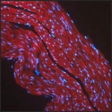
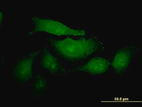
Immunocytochemistry Analysis: 10 µg/mL from a representative lot detected CEACAM1/CD66a on the surface of HT-29 human colorectal carcinoma cells (Courtesy of Dr. B. Singer, University Duisburg-Essen, Germany).
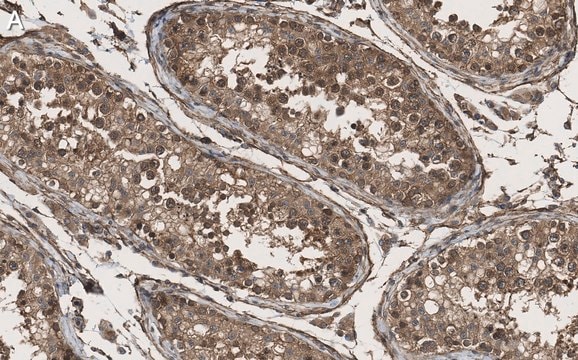
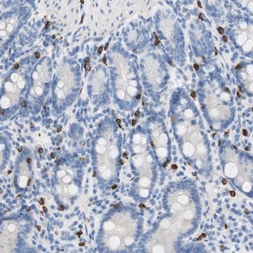
Immunohistochemistry Analysis: 10 µg/mL from a representative lot detected CEACAM1/CD66a immunoreactivity in human jejunum tissue (Courtesy of Dr. B. Singer, University Duisburg-Essen, Germany).
Western Blotting Analysis: 10 µg/mL from a representative lot detected the exogensously expressed human CEACAM1/CD66a extracellular domain-Fc fusion in lysates from transfected cells (Courtesy of Dr. B. Singer, University Duisburg-Essen, Germany).
Flow Cytometry Analysis: Representative lots, either unconjugated or FITC-conjugated, detected CECAM1 immunoreactivity on the surface of human peripheral blood naïve and memory B-cells (Khairnar, V., et al. (2015). Nat. Commun. 6:6217; Seifert, M., et al. (2015). Proc. Natl. Acad. Sci. 112(6):E546-555).
Flow Cytometry Analysis: A representative lot detected CEACAM1/CD66a induction on the surface of normal human bronchial epithelial (NHBE) cells stimulated with poly(I:C) or interferons (Klaile, E., et al. (2013) Respir Res. 14:85).
Flow Cytometry Analysis: A representative lot detected CEACAM1/CD66a expression on the surface of 2 day-starved human epithelial (HT29, T102/3) and endothelial (AS-M.5) cells, as well as multivesicular bodies (MVBs) derived from these cells (Muturi H.T., et al. (2013) PLoS One. 8(9):e74654).
Immunocytochemistry Analysis: A representative lot detected CEACAM1/CD66a immunoreactivity on the surface of normal human bronchial epithelial (NHBE) cells by indirect immunofluorescence staining (Klaile, E., et al. (2013) Respir Res. 14:85).
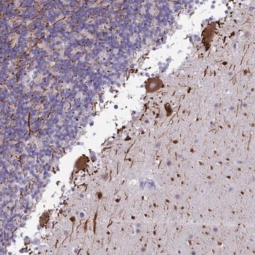
Immunohistochemistry Analysis: A representative lot detected CEACAM1/CD66a in paraffin-embedded human lung cancer tissue sections (Klaile, E., et al. (2013) Respir Res. 14:85).
Western Blot Analysis: A representative lot detected CEACAM1/CD66a in 2 day-starved AS-M.5 human endothelialn cells and AS-M.5-derived multivesicular bodies (MVBs) (Muturi H.T., et al. (2013) PLoS One. 8(9):e74654).
Flow Cytometry Analysis: 10 µg/mL from a representative lot detected the exogenously expressed human CEACAM1/CD66a on the surface of transfected HeLa cells (Courtesy of Dr. B. Singer, University Duisburg-Essen, Germany).
Immunocytochemistry Analysis: 10 µg/mL from a representative lot detected CEACAM1/CD66a on the surface of HT-29 human colorectal carcinoma cells (Courtesy of Dr. B. Singer, University Duisburg-Essen, Germany).
Immunohistochemistry Analysis: 10 µg/mL from a representative lot detected CEACAM1/CD66a immunoreactivity in human jejunum tissue (Courtesy of Dr. B. Singer, University Duisburg-Essen, Germany).
Western Blotting Analysis: 10 µg/mL from a representative lot detected the exogensously expressed human CEACAM1/CD66a extracellular domain-Fc fusion in lysates from transfected cells (Courtesy of Dr. B. Singer, University Duisburg-Essen, Germany).
Flow Cytometry Analysis: Representative lots, either unconjugated or FITC-conjugated, detected CECAM1 immunoreactivity on the surface of human peripheral blood naïve and memory B-cells (Khairnar, V., et al. (2015). Nat. Commun. 6:6217; Seifert, M., et al. (2015). Proc. Natl. Acad. Sci. 112(6):E546-555).
Flow Cytometry Analysis: A representative lot detected CEACAM1/CD66a induction on the surface of normal human bronchial epithelial (NHBE) cells stimulated with poly(I:C) or interferons (Klaile, E., et al. (2013) Respir Res. 14:85).
Flow Cytometry Analysis: A representative lot detected CEACAM1/CD66a expression on the surface of 2 day-starved human epithelial (HT29, T102/3) and endothelial (AS-M.5) cells, as well as multivesicular bodies (MVBs) derived from these cells (Muturi H.T., et al. (2013) PLoS One. 8(9):e74654).
Immunocytochemistry Analysis: A representative lot detected CEACAM1/CD66a immunoreactivity on the surface of normal human bronchial epithelial (NHBE) cells by indirect immunofluorescence staining (Klaile, E., et al. (2013) Respir Res. 14:85).
Immunohistochemistry Analysis: A representative lot detected CEACAM1/CD66a in paraffin-embedded human lung cancer tissue sections (Klaile, E., et al. (2013) Respir Res. 14:85).
Western Blot Analysis: A representative lot detected CEACAM1/CD66a in 2 day-starved AS-M.5 human endothelialn cells and AS-M.5-derived multivesicular bodies (MVBs) (Muturi H.T., et al. (2013) PLoS One. 8(9):e74654).
This Anti-CEACAM1/CD66a Antibody, clone B3-17 is validated for use in ELISA, Flow Cytometry, Immunocytochemistry, Immunohistochemistry, and Western Blotting for the detection of human CEACAM1/CD66a.
품질
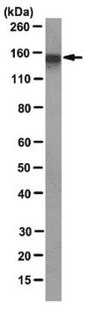
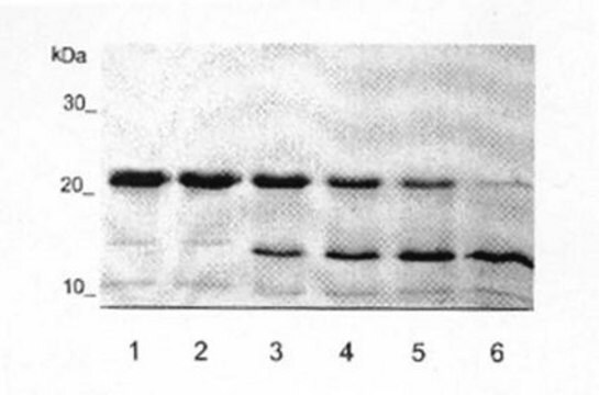
Evaluated by Western Blotting in HepG2 cell lysate.
Western Blotting Analysis: 2 µg/mL of this antibody detected CEACAM1/CD66a in 10 µg of HepG2 cell lysate.
Western Blotting Analysis: 2 µg/mL of this antibody detected CEACAM1/CD66a in 10 µg of HepG2 cell lysate.
표적 설명
~160 kDa observed. Target band size appears larger than the calculated molecular weights of 57.56/53,80 kDa (pro-/mature isoform 1; BGPa, CEACAM1-4L, TM1-CEA), 45.95/42.19 kDa (pro-/mature isoform 2; BGPg, CEACAM1-4C1), 35.30/31.54 kDa (pro-/mature isoform 3; BGPh, CEACAM1-3), 38.58/34.81 kDa (pro-/mature isoform 4; BGPi, CEACAM1-3C2), 50.35/46.58 kDa (pro-/mature isoform 5; BGPy, CEACAM1-3AL), 46.91/43.15 kDa (pro-/mature isoform 6; BGPb, CEACAM1-3L, TM2-CEA), 27.43/23.67 kDa (pro-/mature isoform 7; BGPx, CEACAM1-1L), 50.52/46.76 kDa (pro-/mature isoform 8; BGPc, CEACAM1-4S, TM3-CEA), 43.06/39.30 kDa (pro-/mature isoform 9; BGPz, CEACAM1-3AS), 51.15/47.38 kDa (pro-/mature isoform 10), 39.87/36.11 kDa ((pro-/mature isoform 11; BGPd, CEACAM1-3S) due to glycosylation. Uncharacterized bands may be observed in some lysates.
물리적 형태
Format: Purified
기타 정보
Concentration: Please refer to lot specific datasheet.
적합한 제품을 찾을 수 없으신가요?
당사의 제품 선택기 도구.을(를) 시도해 보세요.
Storage Class Code
12 - Non Combustible Liquids
WGK
WGK 1
Flash Point (°F)
Not applicable
Flash Point (°C)
Not applicable
시험 성적서(COA)
제품의 로트/배치 번호를 입력하여 시험 성적서(COA)을 검색하십시오. 로트 및 배치 번호는 제품 라벨에 있는 ‘로트’ 또는 ‘배치’라는 용어 뒤에서 찾을 수 있습니다.
Marc B Bechmann et al.
Oncotarget, 11(43), 3886-3899 (2020-11-17)
CEACAM5 is overexpressed in many primary breast carcinomas. However, the exact role of CEACAM5 in breast cancer tumorigenesis remains unresolved. Here, we examined a repository of 110 cryopreserved primary breast carcinomas by immunohistochemistry to assess the distribution of CEACAM5 in
활성 필터
자사의 과학자팀은 생명 과학, 재료 과학, 화학 합성, 크로마토그래피, 분석 및 기타 많은 영역을 포함한 모든 과학 분야에 경험이 있습니다..
고객지원팀으로 연락바랍니다.