콘텐츠로 건너뛰기
Merck
모든 사진(2)

문서

MABT339

Sigma-Aldrich

Anti-ZO-1 Antibody, clone 5G6.1

clone 5G6.1, from mouse

동의어(들):

Tight junction protein ZO-1, Tight junction protein 1, Zona occludens protein 1, Zonula occludens protein 1, ZO-1

로그인조직 및 계약 가격 보기


About This Item

UNSPSC 코드:
12352203
eCl@ss:
32160702
NACRES:
NA.41

생물학적 소스

mouse

Quality Level

항체 형태

purified immunoglobulin

항체 생산 유형

primary antibodies

클론

5G6.1, monoclonal

종 반응성

rat, human

기술

immunohistochemistry: suitable (paraffin)
western blot: suitable

동형

IgG2aκ

NCBI 수납 번호

UniProt 수납 번호

배송 상태

wet ice

타겟 번역 후 변형

unmodified

유전자 정보

human ... TJP1(7082)

일반 설명

Tight junction protein ZO-1 (UniProt Q07157; also known as Tight junction protein 1, Zona occludens protein 1, Zonula occludens protein 1) is encoded by the TJP1 gene (also known as ZO1) (Gene ID 7082) in human. The tight junction (TJ) constitutes the barrier between the apical and the basolateral domains of the plasma membrane. The assembly and permeability of this barrier are dependent on the zonula occludens (ZO) membrane-associated guanylate kinase (MAGUK) proteins ZO-1, ZO-2, and ZO-3. ZO-1, a 210-225 kDa protein, is found at the submembranous domain of TJs in epithelia and endothelia. ZO-1 contains three PDZ domains (PDZ1/aa23-109; PDZ2/aa181-261; PDZ3/aa422-502) at its N-treminal end, followed by an SH3 domain (aa519-580), a GUK/GK homology domain (aa632-782), an acidic domain (aa817-894), an alpha spliced domain (aa921-1000), and a C-terminal proline-rich/PR domain. ZO-1 is a phosphoprotein and a known substrate of serine/threonine kinases ZAK and of PKC. MAPK signaling pathway regulates tyrosine phosphorylation of ZO-1, and MEK1 inhibition in Ras transformed epithelial cells is reported to result in tyrosine phosphorylation of ZO-1 and occludin. ZO-1 interacts with claudins, JAM, ZO-2, and ZO-3 through its PDZ domains, while its GK module mediates interaction with occludin. ZO-1 also binds actin cytoskeleton and actin-binding protein 4.1 through its carboxyl terminal end. In addition, ZO-1 associates with AF-6 and cingulin at the TJ, as well as with the adherens junction protein alpha-catenin and with the gap junction proteins connexins 43 and 45.

면역원

GST-tagged recombinant protein corresponding to human ZO-1.

애플리케이션

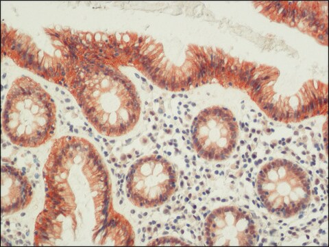
Immunohistochemistry Analysis: A 1:1,000 dilution from a representative lot detected ZO-1 in human and rat kidney tissues.
Research Category
Cell Structure
Research Sub Category
Adhesion (CAMs)
This Anti-ZO-1 Antibody, clone 5G6.1 is validated for use in Western Blotting, Immunocytochemistry, Flow Cytometry, ELISA for the detection of ZO-1 .

품질

Evaluated by Western Blotting in HCT116 cell lysate.

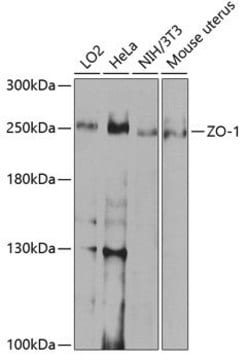
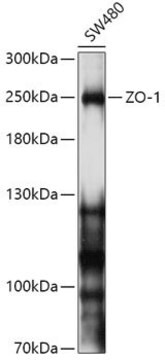
Western Blotting Analysis: 0.5 µg/mL of this antibody detected ZO-1 in 200 µg of HCT116 cell lysate.

표적 설명

~195 kDa observed

물리적 형태

Format: Purified
Protein G Purified
Purified mouse monoclonal IgG2aκ antibody in buffer containing 0.1 M Tris-Glycine (pH 7.4), 150 mM NaCl with 0.05% sodium azide.

저장 및 안정성

Stable for 1 year at 2-8°C from date of receipt.

기타 정보

Concentration: Please refer to lot specific datasheet.

면책조항

Unless otherwise stated in our catalog or other company documentation accompanying the product(s), our products are intended for research use only and are not to be used for any other purpose, which includes but is not limited to, unauthorized commercial uses, in vitro diagnostic uses, ex vivo or in vivo therapeutic uses or any type of consumption or application to humans or animals.

Not finding the right product?  

Try our 제품 선택기 도구.

Storage Class Code

12 - Non Combustible Liquids

WGK

WGK 1

Flash Point (°F)

Not applicable

Flash Point (°C)

Not applicable


시험 성적서(COA)

제품의 로트/배치 번호를 입력하여 시험 성적서(COA)을 검색하십시오. 로트 및 배치 번호는 제품 라벨에 있는 ‘로트’ 또는 ‘배치’라는 용어 뒤에서 찾을 수 있습니다.

이 제품을 이미 가지고 계십니까?

문서 라이브러리에서 최근에 구매한 제품에 대한 문서를 찾아보세요.

문서 라이브러리 방문

Adam M Jorgensen et al.
Tissue engineering. Part A, 26(9-10), 512-526 (2019-12-22)
Over 1 million burn injuries are treated annually in the United States, and current tissue engineered skin fails to meet the need for full-thickness replacement. Bioprinting technology has allowed fabrication of full-thickness skin and has demonstrated the ability to close
Duobin Zhang et al.
Journal of leukocyte biology, 111(5), 953-966 (2021-09-30)
Ischemic stroke (IS) can cause disability and death, and microglia as the immune component of the CNS can release inflammatory factors and participate in blood-brain barrier (BBB) dysfunction. This study aimed to investigate the effects of long noncoding RNA (lncRNA)
Muhammad Ali et al.
Stem cell reports, 16(9), 2320-2335 (2021-08-07)
Here, we evaluate the efficacy of cryopreserved human embryonic stem cell (hESC)-derived corneal endothelial cells (CECs) to form a functional monolayer of corneal endothelium (CE) in rabbits and monkeys. We injected cryopreserved hESC-derived CECs into the anterior chamber of rabbits
Danielle B Buglak et al.
Angiogenesis, 23(4), 567-575 (2020-07-24)
Proper blood vessel formation requires coordinated changes in endothelial cell polarity and rearrangement of cell-cell junctions to form a functional lumen. One important regulator of cell polarity is the centrosome, which acts as a microtubule organizing center. Excess centrosomes perturb

문서

Millicell® hanging cell culture inserts for staining and microscopic analysis directly from the insert. Learn to utilize hanging cell culture inserts in barrier measurements.

관련 콘텐츠

Step-by-step protocol for generating apical-out human gut organoids for microbiome, ADME/Tox, viral and gastrointestinal related disease research. See the complete organoid culture protocol.

Monitor barrier formation using colon PDOs, iPSC-derived colon organoids, Millicell® cell culture inserts, and the Millicell® ERS. 3.0.

자사의 과학자팀은 생명 과학, 재료 과학, 화학 합성, 크로마토그래피, 분석 및 기타 많은 영역을 포함한 모든 과학 분야에 경험이 있습니다..

고객지원팀으로 연락바랍니다.