
가격 및 재고 정보를 현재 이용할 수 없음 고객지원팀으로 연락바랍니다.
추천 제품


생물학적 소스
mouse
Quality Level
항체 형태
purified immunoglobulin
항체 생산 유형
primary antibodies
클론
3H4.3, monoclonal
종 반응성
mouse, rat, human
기술
immunohistochemistry: suitable (paraffin)
western blot: suitable
동형
IgG1κ
NCBI 수납 번호
UniProt 수납 번호
배송 상태
ambient
타겟 번역 후 변형
unmodified
유전자 정보
human ... DLG4(1742)
rat ... Dlg4(29495)
일반 설명
Disks large homolog 4 (UniProt: P31016; also known as Postsynaptic density protein 95, PSD-95, Synapse-associated protein 90, SAP-90, SAP90) is encoded by the Dlg4 (also known as Dlgh4, Psd95) gene (Gene ID: 29495) in rat species. PSD95 belong to the family of membrane-associated guanylate kinases (MAGUKs), which also include PSD-93, SAP97 and SAP102. PSD95 is predominantly expressed in the hippocampus CA1 region and prefrontal cortex of the brain, where it localizes in a somatodendritic pattern in the post-synaptic membrane and in presynaptic axon terminals of inhibitory neurons. PDS95 is the major scaffolding protein in the excitatory postsynaptic density (PSD) and a potent regulator of synaptic strength via interactions with a diverse PSD proteins, including NMDA receptors (NMDARs), AMPA receptor (AMPAR) complexes via Stargazin/TARP, adhesion molecules, as well as other scaffolding proteins, such as GKAP and Shank. PSD95 and neuronal nitric-oxide synthase (nNOS) interaction plays a role in maintaining hypersensitivity in acute and chronic pain. The assembly of the GluR6-PSD95-CaMKII complex is reported to mediate injury induced by brain ischemia by facilitating GluR6 serine phosphorylation by CaMKII and the activation of JNK. Overexpression or depletion of DLG4 changes the ratio of excitatory to inhibitory synapses in hippocampal neurons.
특이성
Clone 3H4.3 detects PSD95 in human and mouse brain.
면역원
KLH-conjugated linear peptide corresponding to 15 amino acids from the N-terminal region of rat PSD95 protein.
애플리케이션
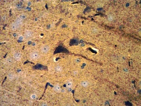
Anti-PSD95, clone 3H4.3, Cat. No. MABN1194, is a highly specific mouse monoclonal antibody that targets PSD95 and has been tested in Immunohistochemistry (Paraffin) and Western Blotting.
Immunohistochemistry Analysis: A 1:50 dilution from a representative lot detected PSD95 in human cerebellum, rat cerebellum, and rat brain stem tissues.
Research Category
Neuroscience
Neuroscience
품질
Evaluated by Western Blotting in mouse brain tissue lysate.
Western Blotting Analysis: 0.5 µg/mL of this antibody detected PSD-95 in 10 µg of mouse brain tissue lysate.
Western Blotting Analysis: 0.5 µg/mL of this antibody detected PSD-95 in 10 µg of mouse brain tissue lysate.
표적 설명
~95 kDa observed; 80.46 kDa calculated. Uncharacterized bands may be observed in some lysate(s).
물리적 형태
Format: Purified
Protein G purified
Purified mouse monoclonal antibody IgG1 in buffer containing 0.1 M Tris-Glycine (pH 7.4), 150 mM NaCl with 0.05% sodium azide.
저장 및 안정성
Stable for 1 year at 2-8°C from date of receipt.
기타 정보
Concentration: Please refer to lot specific datasheet.
면책조항
Unless otherwise stated in our catalog or other company documentation accompanying the product(s), our products are intended for research use only and are not to be used for any other purpose, which includes but is not limited to, unauthorized commercial uses, in vitro diagnostic uses, ex vivo or in vivo therapeutic uses or any type of consumption or application to humans or animals.
적합한 제품을 찾을 수 없으신가요?
당사의 제품 선택기 도구.을(를) 시도해 보세요.
Storage Class Code
12 - Non Combustible Liquids
WGK
WGK 1
시험 성적서(COA)
제품의 로트/배치 번호를 입력하여 시험 성적서(COA)을 검색하십시오. 로트 및 배치 번호는 제품 라벨에 있는 ‘로트’ 또는 ‘배치’라는 용어 뒤에서 찾을 수 있습니다.
Peng Chen et al.
Cell death & disease, 12(4), 403-403 (2021-04-16)
The genes encoding for neuregulin1 (NRG1), a growth factor, and its receptor ErbB4 are both risk factors of major depression disorder and schizophrenia (SZ). They have been implicated in neural development and synaptic plasticity. However, exactly how NRG1 variations lead
Shatrunjai Giri et al.
Journal of neuroscience research, 99(7), 1815-1834 (2021-04-06)
Rapid eye movement sleep (REMS) favors brain development and memory, while it is decreased in neurodegenerative diseases. REMS deprivation (REMSD) affects several physiological processes including memory consolidation; however, its detailed mechanism(s) of action was unknown. REMS reduces, while REMSD elevates
활성 필터
자사의 과학자팀은 생명 과학, 재료 과학, 화학 합성, 크로마토그래피, 분석 및 기타 많은 영역을 포함한 모든 과학 분야에 경험이 있습니다..
고객지원팀으로 연락바랍니다.