
추천 제품


생물학적 소스
mouse
Quality Level
항체 형태
purified antibody
항체 생산 유형
primary antibodies
클론
8E8, monoclonal
종 반응성
mouse
종 반응성(상동성에 의해 예측)
human (based on 100% sequence homology)
기술
activity assay: suitable
flow cytometry: suitable
동형
IgG2bκ
NCBI 수납 번호
UniProt 수납 번호
배송 상태
wet ice
타겟 번역 후 변형
unmodified
유전자 정보
human ... PARD3(56288)
일반 설명
PAR-3 (also known as Coagulation factor II receptor-like 2, Thrombin receptor-like 2 or F2RL2) is an adapter protein involved in asymmetrical cell division and cell polarization processes. It seems to play a central role in the formation of epithelial tight junctions. Its association with PARD6B may prevent the interaction of PAR-3 with F11R/JAM1, thereby preventing tight junction assembly. The PARD6-PAR-3 complex links GTP bound Rho small GTPases to atypical protein kinase C proteins. PAR-3 interacts with PARD6A and PARD6B. Isoform 2, but not at least isoform 3 interacts with PRKCZ. PAR-3 interacts with PRCKI. PAR-3 forms part of a complex with PARD6A or PARD6B, PRKCI or PRKCZ and CDC42 or RAC1. PAR-3 interacts with F11R/JAM1. Antibodies against PAR-3 are present in sera from patients with cutaneous T cell lymphomas.
면역원
KLH-conjugated linear peptide corresponding to human PAR-3.
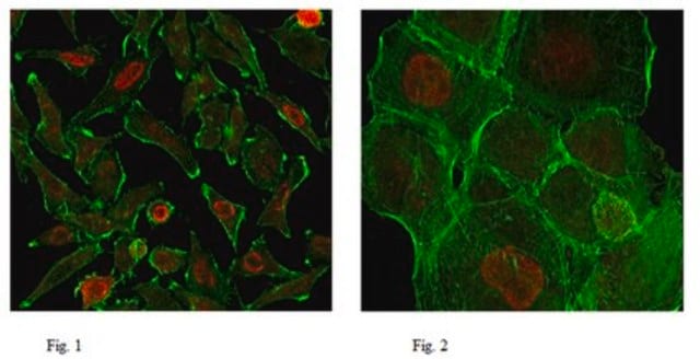
애플리케이션
Anti-PAR-3 Antibody, clone 8E8 detects level of PAR-3 & has been published & validated for use in FC, EA.
Flow Cytometry Analysis: A previous lot was used by an independent laboratory in FC. (Petrova, Y., et al. (2008). Centr Eur J Immunol. 33 (1):14-18.)
Activity Assay Analysis (platelet aggregation): A previous lot was used by an independent laboratory in platelet aggregation assay. (Petrova, Y., et al. (2008). Centr Eur J Immunol. 33 (1):14-18.)
Activity Assay Analysis (platelet aggregation): A previous lot was used by an independent laboratory in platelet aggregation assay. (Petrova, Y., et al. (2008). Centr Eur J Immunol. 33 (1):14-18.)
Research Category
Infectious Diseases
Infectious Diseases
Research Sub Category
Inflammation & Autoimmune Mechanisms
Inflammation & Autoimmune Mechanisms
품질
Evaluated by Flow Cytometry in mouse platelets from washed whole blood (heparinized).
Flow Cytometry Analysis: 2 µg of this antibody detected PAR-3 in mouse platelets from washed whole blood (heparinized).
Flow Cytometry Analysis: 2 µg of this antibody detected PAR-3 in mouse platelets from washed whole blood (heparinized).
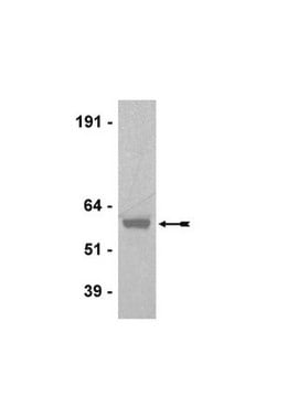
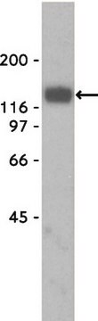
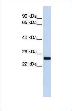
표적 설명
40 kDa calculated
결합
Replaces: MABS174
물리적 형태
Format: Purified
Protein G Purified
Purified mouse monoclonal IgG2bκ in buffer containing 0.1 M Tris-Glycine (pH 7.4), 150 mM NaCl with 0.05% sodium azide.
저장 및 안정성
Stable for 1 year at 2-8°C from date of receipt.
분석 메모
Control
Mouse platelets from washed whole blood (heparinized)
Mouse platelets from washed whole blood (heparinized)
기타 정보
Concentration: Please refer to the Certificate of Analysis for the lot-specific concentration.
면책조항
Unless otherwise stated in our catalog or other company documentation accompanying the product(s), our products are intended for research use only and are not to be used for any other purpose, which includes but is not limited to, unauthorized commercial uses, in vitro diagnostic uses, ex vivo or in vivo therapeutic uses or any type of consumption or application to humans or animals.
적합한 제품을 찾을 수 없으신가요?
당사의 제품 선택기 도구.을(를) 시도해 보세요.
Storage Class Code
12 - Non Combustible Liquids
WGK
WGK 1
Flash Point (°F)
Not applicable
Flash Point (°C)
Not applicable
시험 성적서(COA)
제품의 로트/배치 번호를 입력하여 시험 성적서(COA)을 검색하십시오. 로트 및 배치 번호는 제품 라벨에 있는 ‘로트’ 또는 ‘배치’라는 용어 뒤에서 찾을 수 있습니다.
Hiroyuki Nakajima et al.
Journal of cell science, 123(Pt 4), 555-566 (2010-01-28)
Cell-shape change in epithelial structures is fundamental to animal morphogenesis. Recent studies identified myosin-II as the major generator of driving forces for cell-shape changes during morphogenesis. Lulu (Epb41l5) is a major regulator of morphogenesis, although the downstream molecular and cellular
Michael L Drummond et al.
The Journal of cell biology, 217(9), 3255-3266 (2018-06-28)
Primary cilia are polarized organelles that allow detection of extracellular signals such as Hedgehog (Hh). How the cytoskeleton supporting the cilium generates and maintains a structure that finely tunes cellular response remains unclear. Here, we find that regulation of actin
Virginia Fernández et al.
The EMBO journal, 39(21), e105479-e105479 (2020-09-29)
Structural integrity and cellular homeostasis of the embryonic stem cell niche are critical for normal tissue development. In the telencephalic neuroepithelium, this is controlled in part by cell adhesion molecules and regulators of progenitor cell lineage, but the specific orchestration
Kaviya Chinnappa et al.
Science advances, 8(2), eabj4010-eabj4010 (2022-01-13)
The evolutionary expansion and folding of the mammalian cerebral cortex resulted from amplification of progenitor cells during embryonic development. This process was reversed in the rodent lineage after splitting from primates, leading to smaller and smooth brains. Genetic mechanisms underlying
활성 필터
자사의 과학자팀은 생명 과학, 재료 과학, 화학 합성, 크로마토그래피, 분석 및 기타 많은 영역을 포함한 모든 과학 분야에 경험이 있습니다..
고객지원팀으로 연락바랍니다.