
가격 및 재고 정보를 현재 이용할 수 없음
추천 제품


생물학적 소스
mouse
Quality Level
항체 형태
purified immunoglobulin
항체 생산 유형
primary antibodies
클론
29E.2A3.C6, monoclonal
종 반응성
rat, chimpanzee, human
기술
flow cytometry: suitable
immunocytochemistry: suitable
immunohistochemistry: suitable (paraffin)
neutralization: suitable
동형
IgG2bκ
NCBI 수납 번호
UniProt 수납 번호
배송 상태
dry ice
타겟 번역 후 변형
unmodified
유전자 정보
human ... CD274(29126)
일반 설명
Programmed cell death 1 ligand 1 (UniProt Q9NZQ7; also known as B7-H1, B7 homolog 1, CD274, PD-L1, PDCD1 ligand 1, Programmed death ligand 1) is encoded by the CD274 (also known as B7H1, PDCD1L1, PDCD1LG1, PDL1) gene (Gene ID 29126) in human. PD-1 and PD-1 ligands 1&2 (PD-L1 and PD-L2) are B7:CD28 family members that regulate T cell activation and peripheral tolerance. When engaged together with the TCR, the interaction of PD-1 with its ligands delivers an inhibitory signal to T cell proliferation and cytokine production. While PD-L1 is broadly expressed in hematopoietic and nonhematopoietic cells, PD-L2 expression is highly restricted to antigen presenting cells (APCs), including dendritic cells (DCs) and macrophages. The PD-1 pathway plays a key role in the progressive loss of effector T cell responses during chronic HIV infection. Under some conditions, blockade of this pathway is able to restore many T cell functions. PD-L1 is initially produced with signal peptide (a.a. 1-18) sequence, the removal of which yields the mature protien with a large extracellular (a.a. 19-238) region that contains an Ig-like V-type domain (a.a. 19-127) and an Ig-like C2-type domain (a.a. 133-225), followed by a transmembrane domain (a.a. 239-259) and a cytoplasmic tail (a.a. 260-290).
특이성
Clone 29E.2A3.C6 immunostained the surface of 300.19 murine pre-B lymphoma cells transfected with human PD-L1, but not the untransfected cells or cells transfected with human PD-L2 (Brown, J.A., et al. (2003). J. Immunol. 170(3):1257-1266). Reactivity toward spliced isoform 2 (PD-L1II) and isoform 3 has not been determined.
면역원
Epitope: Extracellular domain.
Recombinant full-length human PD-L1.
애플리케이션
Anti-PD-L1 Antibody, clone 29E.2A3.C6, Azide Free is an antibody against PD-L1 for use in Immunohistochemistry (Paraffin), Flow Cytometry, Neutralizing, Immunocytochemistry.
Immunohistochemistry Analysis: An 1:50 dilution of this antibody from a representative lot detected PD-L1 in human kidney and rat colon tissue.
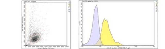
Flow Cytometry Analysis: 0.1 µg of this antibody from a representative lot detected PD-L1 in MDA-MB-231 cells.
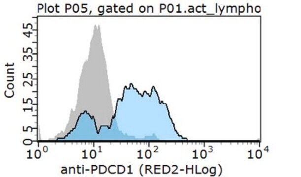
Flow Cytometry Analysis: A representative lot immunostained the surface of 300.19 murine pre-B lymphoma cells transfected with human PD-L1, but not the untransfected cells or cells transfected with human PD-L2 (Brown, J.A., et al. (2003). J. Immunol. 170(3):1257-1266).
Flow Cytometry Analysis: A representative lot detected surface PD-L1 expression on human breast cancer cell lines MDA-231, SKBR-3, and MCF-7, but not BT-474 (Latchman, Y., et al. (2001). J. Nat. Immunol.2(3):261-268).
Immunohistochemistry Analysis: A representative lot detected PD-L1 expression pattern in frozen fetal (thymus & cardiac tissue) and paraffin-embedded adult (tonsillar germinal center and placenta) human tissue sections (Brown, J.A., et al. (2003). J. Immunol. 170(3):1257-1266).
Immunohistochemistry Analysis: A representative lot detected PD-L1 expression in paraffin-embedded human cancer tissue sections, including anaplastic large cell lymphoma, squamous cell carcinoma of the tongue, colon adenocarcinoma, and invasive breast ductal carcinoma (Brown, J.A., et al. (2003). J. Immunol. 170(3):1257-1266).
Neutralizing Analysis: A representative lot competed against human PD-1 extracellular domain Ig fusion for the binding of exogenously expressed human PD-L1 on the surface of 300.19 murine pre-B lymphoma cells (Brown, J.A., et al. (2003). J. Immunol. 170(3):1257-1266).
Neutralizing Analysis: A representative lot enhanced cytokines production from CD4 T-cells upon HIV Gag peptides stimulation of CD8+-depleted human PBMCs (Porichis, F., et al. (2011). Blood. 118(4):965-974).
Neutralizing Analysis: A representative lot restored HCV peptides-induced expansion of CD8+ T cells in cultured intrahepatic lymphocytes from a chimpanzee with 10 years of chronic HCV infection (Fuller, M.J., et al. (2013). Proc. Natl. Acad. Sci. U. S. A. 110(37):15001-15006).
Neutralizing Analysis: Dual blockage of both DP-L1 clone 29E.2A3.C6 Fab fragment and DP-L2 by clone 24F.10C12 (Cat. No. MABC969) Fab fragment on PBMC-derived dendric cells (DCs) boosted the enhancing effect on CD4+ T-cell proliferation and cytokine release seen with DP-L2 blockage alone in allogenic cultures with immature DCs (iDC), mature DCs (mDC), and IL-10-treated mDCs (Brown, J.A., et al. (2003). J. Immunol. 170(3):1257-1266).
Immunocytochemistry Analysis: A representative lot detected an upregulated PD-L1 immunoreactivity in human CD14+ monocyte-derived macrophages (MDMs) following HIV infection or TLR4/8 stimulation (by LPS or CL097 treatment) by fluorescent immunocytochemistry using paraformaldehyde-fixed MDMs (Rodríguez-García, M., et al. (2011). J. Leukoc. Biol. 89(4) 507–515).
Flow Cytometry Analysis: 0.1 µg of this antibody from a representative lot detected PD-L1 in MDA-MB-231 cells.
Flow Cytometry Analysis: A representative lot immunostained the surface of 300.19 murine pre-B lymphoma cells transfected with human PD-L1, but not the untransfected cells or cells transfected with human PD-L2 (Brown, J.A., et al. (2003). J. Immunol. 170(3):1257-1266).
Flow Cytometry Analysis: A representative lot detected surface PD-L1 expression on human breast cancer cell lines MDA-231, SKBR-3, and MCF-7, but not BT-474 (Latchman, Y., et al. (2001). J. Nat. Immunol.2(3):261-268).
Immunohistochemistry Analysis: A representative lot detected PD-L1 expression pattern in frozen fetal (thymus & cardiac tissue) and paraffin-embedded adult (tonsillar germinal center and placenta) human tissue sections (Brown, J.A., et al. (2003). J. Immunol. 170(3):1257-1266).
Immunohistochemistry Analysis: A representative lot detected PD-L1 expression in paraffin-embedded human cancer tissue sections, including anaplastic large cell lymphoma, squamous cell carcinoma of the tongue, colon adenocarcinoma, and invasive breast ductal carcinoma (Brown, J.A., et al. (2003). J. Immunol. 170(3):1257-1266).
Neutralizing Analysis: A representative lot competed against human PD-1 extracellular domain Ig fusion for the binding of exogenously expressed human PD-L1 on the surface of 300.19 murine pre-B lymphoma cells (Brown, J.A., et al. (2003). J. Immunol. 170(3):1257-1266).
Neutralizing Analysis: A representative lot enhanced cytokines production from CD4 T-cells upon HIV Gag peptides stimulation of CD8+-depleted human PBMCs (Porichis, F., et al. (2011). Blood. 118(4):965-974).
Neutralizing Analysis: A representative lot restored HCV peptides-induced expansion of CD8+ T cells in cultured intrahepatic lymphocytes from a chimpanzee with 10 years of chronic HCV infection (Fuller, M.J., et al. (2013). Proc. Natl. Acad. Sci. U. S. A. 110(37):15001-15006).
Neutralizing Analysis: Dual blockage of both DP-L1 clone 29E.2A3.C6 Fab fragment and DP-L2 by clone 24F.10C12 (Cat. No. MABC969) Fab fragment on PBMC-derived dendric cells (DCs) boosted the enhancing effect on CD4+ T-cell proliferation and cytokine release seen with DP-L2 blockage alone in allogenic cultures with immature DCs (iDC), mature DCs (mDC), and IL-10-treated mDCs (Brown, J.A., et al. (2003). J. Immunol. 170(3):1257-1266).
Immunocytochemistry Analysis: A representative lot detected an upregulated PD-L1 immunoreactivity in human CD14+ monocyte-derived macrophages (MDMs) following HIV infection or TLR4/8 stimulation (by LPS or CL097 treatment) by fluorescent immunocytochemistry using paraformaldehyde-fixed MDMs (Rodríguez-García, M., et al. (2011). J. Leukoc. Biol. 89(4) 507–515).
Research Category
Apoptosis & Cancer
Apoptosis & Cancer
Research Sub Category
Apoptosis - Additional
Apoptosis - Additional
품질
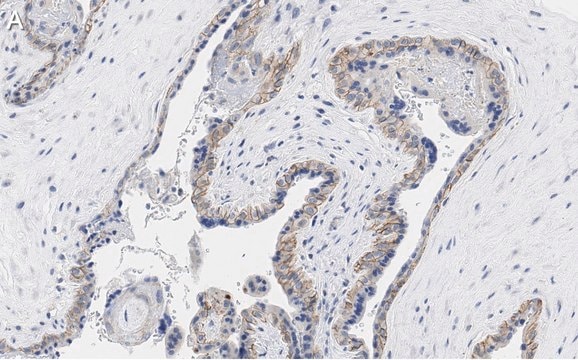
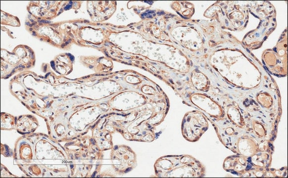
Evaluated by Immunohistochemistry in human cerebral cortex tissue.
Immunohistochemistry Analysis: An 1:50 dilution of this antibody detected PD-L1 in human cerebral cortex tissue.
Immunohistochemistry Analysis: An 1:50 dilution of this antibody detected PD-L1 in human cerebral cortex tissue.
표적 설명
31.02 kDa (isoform 1; PD-L1I), 17.99 kDa (isoform 2; PD-L1II), 18.20 kDa (isoform 3) calculated.
물리적 형태
Format: Purified
Protein G Purified
Purified mouse monoclonal IgG2bκ antibody in PBS without preservatives.
저장 및 안정성
Stable for 1 year at -20°C from date of receipt.
Handling Recommendations: Upon receipt and prior to removing the cap, centrifuge the vial and gently mix the solution. Aliquot into microcentrifuge tubes and store at -20°C. Avoid repeated freeze/thaw cycles, which may damage IgG and affect product performance.
Handling Recommendations: Upon receipt and prior to removing the cap, centrifuge the vial and gently mix the solution. Aliquot into microcentrifuge tubes and store at -20°C. Avoid repeated freeze/thaw cycles, which may damage IgG and affect product performance.
기타 정보
Concentration: Please refer to lot specific datasheet.
면책조항
Unless otherwise stated in our catalog or other company documentation accompanying the product(s), our products are intended for research use only and are not to be used for any other purpose, which includes but is not limited to, unauthorized commercial uses, in vitro diagnostic uses, ex vivo or in vivo therapeutic uses or any type of consumption or application to humans or animals.
적합한 제품을 찾을 수 없으신가요?
당사의 제품 선택기 도구.을(를) 시도해 보세요.
Storage Class Code
12 - Non Combustible Liquids
WGK
WGK 2
Flash Point (°F)
Not applicable
Flash Point (°C)
Not applicable
시험 성적서(COA)
제품의 로트/배치 번호를 입력하여 시험 성적서(COA)을 검색하십시오. 로트 및 배치 번호는 제품 라벨에 있는 ‘로트’ 또는 ‘배치’라는 용어 뒤에서 찾을 수 있습니다.
활성 필터
자사의 과학자팀은 생명 과학, 재료 과학, 화학 합성, 크로마토그래피, 분석 및 기타 많은 영역을 포함한 모든 과학 분야에 경험이 있습니다..
고객지원팀으로 연락바랍니다.