
추천 제품


생물학적 소스
mouse
Quality Level
항체 형태
purified immunoglobulin
항체 생산 유형
primary antibodies
클론
6F-H2, monoclonal
종 반응성
mouse, human
기술
immunocytochemistry: suitable
immunofluorescence: suitable
immunohistochemistry: suitable (paraffin)
immunoprecipitation (IP): suitable
western blot: suitable
동형
IgG1κ
NCBI 수납 번호
UniProt 수납 번호
배송 상태
wet ice
타겟 번역 후 변형
unmodified
유전자 정보
human ... WT1(7490)
일반 설명
Wilms tumor protein (UniProt P19544; also known as WT33) is encoded by the WT1 (also known as DDS, FS, MEACHS, MESOM, NPHS4, WAGR) gene (Gene ID 7490) in human. The Wilms’ tumor gene WT1 was originally identified in the childhood kidney cancer Wilms’ tumor. The N-terminal region of WT1 protein contains a proline-rich region (a.a. 27-83) involved in transcriptional regulation, self-association, and RNA recognition, while its C-terminal region contains four zinc fingers (a.a 323-347, 353-377, 383-405, 414-438) that mediate DNA and RNA binding. The zinc finger domain of WT1 can bind to GC-rich sequences, such as the EGR-1 consensus sequence (5’-GCG(T/G)GGGCG-3’), the WTE motif (5′-GCGTGGGAGT-3′), or (TCC)n motif. Many genes responsible for cell growth and apoptosis, such as Bcl-2, Bcl-xL, BFL1, and c-myc, have been identified as downstream targets of WT1. There are four major alternatively spliced WT1 isoforms resulting from splicing at either or both of exon 5 (17AA) and exon 9 (KTS). All four major WT1 isoforms are overexpressed in leukemia and solid tumors and play oncogenic roles such as inhibition of apoptosis, and promotion of cell proliferation, migration and invasion.
애플리케이션
Immunohistochemistry Analysis: An 1:250 dilution from a representative lot detected Wilms tumor protein in human kidney tissue.
Immunoprecipitation Analysis: A representative lot co-immunoprecipitated CRE-binding protein/CBP together with Wilms tumor protein WT1 from the lysate of a T-SV40 immortalized human glomerular epithelial cell (HGEC) line (Drossopoulou, G.I., et al. (2009). Am. J. Physiol. Renal Physiol. 297(3):F594-F603).
Western Blotting Analysis: A representative lot detected Wilms tumor protein WT1 in the CRE-binding protein/CBP immunoprecipitate obtained from the lysate of a T-SV40 immortalized human glomerular epithelial cell (HGEC) line (Drossopoulou, G.I., et al. (2009). Am. J. Physiol. Renal Physiol. 297(3):F594-F603).
Western Blotting Analysis: A representative lot detected Wilms tumor protein WT1 in lysates from mouse E15.5 embryonic kidney and human melanoma cell lines A375, SK-MEL-28, and WM-266-4 (Wagner, N., et al. (2008). Pflugers Arch. Eur. J. Physiol. 455(5):839-847).
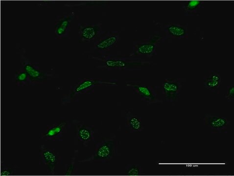
Immunocytochemistry Analysis: A representative lot immunostained the nucleus of methanol-fixed human melanoma A375 cells by fluorescent immunocytochemistry (Wagner, N., et al. (2008). Pflugers Arch. Eur. J. Physiol. 455(5):839-847).
Immunofluorescence Analysis: A representative lot immunostained the PCNA-positive nuclei of proliferating cells in formalin-fixed, paraffin-embedded human melanoma tissue sections by fluorescent immunohistochemistry (Wagner, N., et al. (2008). Pflugers Arch. Eur. J. Physiol. 455(5):839-847).
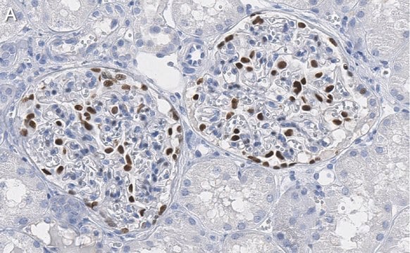
Immunohistochemistry Analysis: A representative lot immunostained glomeruli in formalin-fixed, paraffin-embedded normal human kidney and Wilms′ tumor sections (Wagner, N., et al. (2008). Pediatr. Nephrol. 23(9):1445-1453).
Immunohistochemistry Analysis: A representative lot detected vascular WT1 expression in 95% of 113 paraffin-embedded tumour tissues of various types. In most cases, nuclear WT1 staining of endothelial cells was seen (Wagner, N., et al. (2008). Oncogene. 27(26):3662-3672).
Immunohistochemistry Analysis: A representative lot immunostained the nucleus of perifollicular fibroblasts at the hair follicle in formalin-fixed, paraffin-embedded normal human skin sections. Most common melanocytic nevi do not express WT1, whereas Spitz nevi and dysplastic nevi show cytoplasmic WT1 staining. (Wagner, N., et al. (2008). Pflugers Arch. Eur. J. Physiol. 455(5):839-847).
Immunoprecipitation Analysis: A representative lot co-immunoprecipitated CRE-binding protein/CBP together with Wilms tumor protein WT1 from the lysate of a T-SV40 immortalized human glomerular epithelial cell (HGEC) line (Drossopoulou, G.I., et al. (2009). Am. J. Physiol. Renal Physiol. 297(3):F594-F603).
Western Blotting Analysis: A representative lot detected Wilms tumor protein WT1 in the CRE-binding protein/CBP immunoprecipitate obtained from the lysate of a T-SV40 immortalized human glomerular epithelial cell (HGEC) line (Drossopoulou, G.I., et al. (2009). Am. J. Physiol. Renal Physiol. 297(3):F594-F603).
Western Blotting Analysis: A representative lot detected Wilms tumor protein WT1 in lysates from mouse E15.5 embryonic kidney and human melanoma cell lines A375, SK-MEL-28, and WM-266-4 (Wagner, N., et al. (2008). Pflugers Arch. Eur. J. Physiol. 455(5):839-847).
Immunocytochemistry Analysis: A representative lot immunostained the nucleus of methanol-fixed human melanoma A375 cells by fluorescent immunocytochemistry (Wagner, N., et al. (2008). Pflugers Arch. Eur. J. Physiol. 455(5):839-847).
Immunofluorescence Analysis: A representative lot immunostained the PCNA-positive nuclei of proliferating cells in formalin-fixed, paraffin-embedded human melanoma tissue sections by fluorescent immunohistochemistry (Wagner, N., et al. (2008). Pflugers Arch. Eur. J. Physiol. 455(5):839-847).
Immunohistochemistry Analysis: A representative lot immunostained glomeruli in formalin-fixed, paraffin-embedded normal human kidney and Wilms′ tumor sections (Wagner, N., et al. (2008). Pediatr. Nephrol. 23(9):1445-1453).
Immunohistochemistry Analysis: A representative lot detected vascular WT1 expression in 95% of 113 paraffin-embedded tumour tissues of various types. In most cases, nuclear WT1 staining of endothelial cells was seen (Wagner, N., et al. (2008). Oncogene. 27(26):3662-3672).
Immunohistochemistry Analysis: A representative lot immunostained the nucleus of perifollicular fibroblasts at the hair follicle in formalin-fixed, paraffin-embedded normal human skin sections. Most common melanocytic nevi do not express WT1, whereas Spitz nevi and dysplastic nevi show cytoplasmic WT1 staining. (Wagner, N., et al. (2008). Pflugers Arch. Eur. J. Physiol. 455(5):839-847).
This Anti-Wilms′ Tumor Antibody, NT clone 6F-H2, Ascites Free is validated for use in Immunocytochemistry, Immunoprecipitation, Immunofluorescence, Immunohistochemistry (Paraffin), and Western Blotting for the detection of Wilms′ tumor protein.
품질
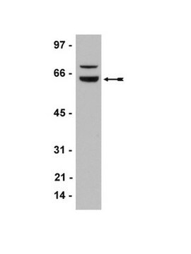
Evaluated by Western Blotting in Jurkat cell lysate.
Western Blotting Analysis: 1.0 µg/mL of this antibody detected Wilms tumor protein in 10 µg of Jurkat cell lysate.
Western Blotting Analysis: 1.0 µg/mL of this antibody detected Wilms tumor protein in 10 µg of Jurkat cell lysate.
표적 설명
~52 kDa observed. 49.19 kDa (isoform 1), 47.20 kDa (isoform 2), 47.51 kDa (isoform 3), 48.87 kDa (isoform 4), 34.45 kDa (isoform 5), 56.88 kDa (isoform 6), 55.21 kDa (isoform 7), 33.09 kDa (isoform 8) calculated. Uncharacterized band(s) may appear in some lysates.
물리적 형태
Format: Purified
기타 정보
Concentration: Please refer to lot specific datasheet.
적합한 제품을 찾을 수 없으신가요?
당사의 제품 선택기 도구.을(를) 시도해 보세요.
Storage Class Code
12 - Non Combustible Liquids
WGK
WGK 1
Flash Point (°F)
Not applicable
Flash Point (°C)
Not applicable
시험 성적서(COA)
제품의 로트/배치 번호를 입력하여 시험 성적서(COA)을 검색하십시오. 로트 및 배치 번호는 제품 라벨에 있는 ‘로트’ 또는 ‘배치’라는 용어 뒤에서 찾을 수 있습니다.
활성 필터
자사의 과학자팀은 생명 과학, 재료 과학, 화학 합성, 크로마토그래피, 분석 및 기타 많은 영역을 포함한 모든 과학 분야에 경험이 있습니다..
고객지원팀으로 연락바랍니다.