추천 제품
생물학적 소스
mouse
Quality Level
100
300
항체 형태
purified antibody
항체 생산 유형
primary antibodies
클론
Lu5, monoclonal
종 반응성(상동성에 의해 예측)
mammals
제조업체/상표
Chemicon®
기술
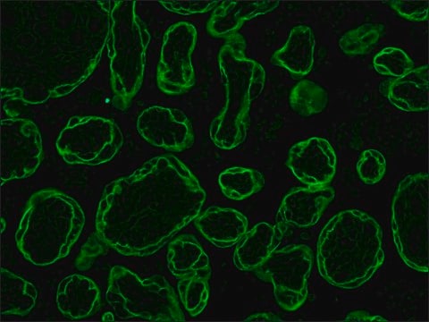
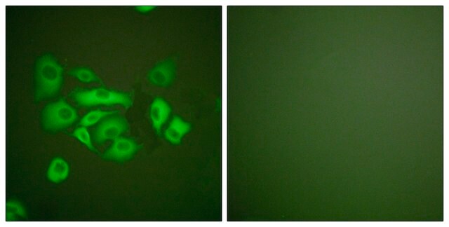
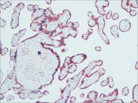
immunohistochemistry: suitable
동형
IgG1
배송 상태
wet ice
특이성
The antibody reacts with an epitope which is common to all cytokeratins respectively with a cytokeratin-associated protein. The antigen recognized by anti-cytokeratin pan is present in all types of epithelia. The antibody reacts with epithelial and mesothelial cells. Exception: Secretory cells are not stained and acinus cells, for example, show only weak staining. The antibody does not react with connective tissue, blood or lymphatic vessels, smooth or skeletal muscles. Exception: When higher antibody concentrations are used, weakly positive reactions can be observed with the smooth muscles of the myometrium, the prostata as well as the gastric muscularis mucosa.The antibodies can be used for differential diagnosis of epithelial tumors, distinction from mesenchymal, neural tumors as well as large-cell lymphomas. Anti-cytokeratin pan stains all epithelial tumors (primary tumors as well as metastases). Exception: Only partially positive staining is observed with theca cell tumors, granulosa cell tumors, adrenal cortex tumors. In mixed tumors (e.g. carcinosarkoma, thymoma, adenolymphoma) only the epithelial regions are stained. Tumors of nonepithelial origin do not react with the antibody. Exception: With high antibody concentrations a weak staining of leiomyosarcoma of the uterus is observed.
면역원
Lung tumor cell lines A549 and A2182.
애플리케이션
Immunohistochemistry: 10-20 μg/mL Optimal working dilutions must be determined by end user.
Tumor Characterization
For tumor characterization a two-step experimental assay is recommended:
1. Characterization of the tissue origin of a tumor with the aid of anti-cytokeratin pan, anti-desmin, anti-glial fibrillary acidic protein, anti-neurofilament and anti-vimentin.
2. Characterization of an epithelial tumor using antibodies to cytokeratin classes which are characteristic for different tissues, e.g. anti-cytokeratin No. 7, anti-cytokeratin No. 18. Sample material: Normal tissue, tumor tissue following surgery or up to 24 h after autopsy, frozen sections or formaldehyde-fixed paraffin embedded sections,
Detection method
Immunohistochemical analysis (detection of the primary antibody e. g. by anti-mouse Ig-peroxidase or anti-mouse Ig-FITC).
Procedure:
Ideal frozen sections are obtained from shock-frozen tissue samples. The frozen sections are air-dried and then fixed with acetone for 5-10 min at -15 to -25°C. Excess acetone is allowed to evaporate at 15-25°C. Material fixed in alcohol can also be used.
Cytocentrifuge preparations of single cells or cell smears are also fixed in acetone. These preparations should, however, not be air dried, the excess acetone is removed by briefly washing in phosphate-buffered saline (PBS). Further treatment as follows:
• Overlay the preparation with 10 - 20 μL antibody solution and incubate in a humid chamber for 30 - 60 min at 15-25°C.
• Dip the slide briefly in PBS and then wash in PBS 3 times for 3 min each (using a fresh PBS bath in each case).
• Wipe the margins of the preparation dry, overlay the preparation with 10 - 20 μL of a solution of anti-mouse lg-FITC or anti-mouse Ig-peroxidase and allow to incubate in a humid chamber for 30 min at 15-25°C.
• Wash the slide in PBS as described above.
The preparation must not be allowed to dry out during any of the steps. lf using an indirect immunofluorescence technique, the preparation should be overlaid with a suitable embedding medium (e. g. Mowiol, Hoechst) and examined under the fluorescence microscope. lf a POD-conjugate is used as the secondary antibody, the preparation should be overlaid with a substrate solution (see below) and incubated at 15-25°C until a clearly visible redbrown color develops. A negative control (e.g. only the labeled secondary antibody) should remain unchanged in color during this incubation period.
Subsequently, the substrate is washed off with PBS and the preparation is stained with hemalum stain for about 1 min. The hemalum solution is washed off with PBS, the preparation is embedded and examined.
Substrate solutions:
Aminoethyl-carbazole: Dissolve 2 mg 3-amino-9-ethylcarbazole with 1.2 mL dimethylsulfoxide and add 28.8 mL Tris-HCI, 0.05 M; pH 7.3; and 20 μL 3% H 2 O 2 , (w/v). Prepare solution freshly each day. Diaminobenzidine: Dissolve 25 mg 3, 3′-diaminobenzidine with 50 mL Tris-HCI, 0.05 M; pH 7.3; and add 40 μL 3% H2O2 , (w/v). Prepare solution freshly each day. Note The antibody solutions have to be absolutely free of precipitate! lf necessary, centrifuge the solutions at high speed prior to use.
This Anti-Cytokeratin Pan Antibody, clone Lu5 is validated for use in IH for the detection of Cytokeratin Pan.
물리적 형태
Format: Purified
Liquid. Buffer = 0.02 M Phosphate buffer, 0.25M NaCl with 0.1% sodium azide.
저장 및 안정성
The lyophilized antibody is stabe when stored at 2-8°C.The reconstituted antibody solution is stable at 2-8°C for three monthes. The solution can be stored in aliquots at -15 to -25 °C for 12 monthes. Avoid repeat freeze/thaw cycles.
기타 정보
Concentration: Please refer to the Certificate of Analysis for the lot-specific concentration.
법적 정보
CHEMICON is a registered trademark of Merck KGaA, Darmstadt, Germany
적합한 제품을 찾을 수 없으신가요?
당사의 제품 선택기 도구.을(를) 시도해 보세요.
Storage Class Code
10 - Combustible liquids
WGK
WGK 2
Flash Point (°F)
Not applicable
Flash Point (°C)
Not applicable
시험 성적서(COA)
제품의 로트/배치 번호를 입력하여 시험 성적서(COA)을 검색하십시오. 로트 및 배치 번호는 제품 라벨에 있는 ‘로트’ 또는 ‘배치’라는 용어 뒤에서 찾을 수 있습니다.
Bhairavi Bhatia et al.
Experimental eye research, 93(6), 852-861 (2011-10-13)
Much controversy has arisen on the nature and sources of stem cells in the adult human retina. Whilst ciliary epithelium has been thought to constitute a source of neural stem cells, a population of Müller glia in the neural retina
Z Zhang et al.
Neuroscience, 174, 10-25 (2010-12-01)
In adult cortices, the ratio of excitatory and inhibitory conductances (E/I ratio) is presumably balanced across a wide range of stimulus conditions. However, it is unknown how the E/I ratio is postnatally regulated, when the strength of synapses are rapidly
자사의 과학자팀은 생명 과학, 재료 과학, 화학 합성, 크로마토그래피, 분석 및 기타 많은 영역을 포함한 모든 과학 분야에 경험이 있습니다..
고객지원팀으로 연락바랍니다.


