추천 제품
생물학적 소스
mouse
Quality Level
항체 형태
purified antibody
항체 생산 유형
primary antibodies
클론
GNS3 (8A5D12), monoclonal
종 반응성
human, mouse
포장
antibody small pack of 25 μg
제조업체/상표
Upstate®
기술
immunohistochemistry: suitable
immunoprecipitation (IP): suitable
western blot: suitable
동형
IgG
NCBI 수납 번호
UniProt 수납 번호
배송 상태
dry ice
타겟 번역 후 변형
unmodified
유전자 정보
human ... CCNB1(891)
mouse ... Ccnb1(268697)
관련 카테고리
일반 설명
G2/mitotic-specific cyclin-B1 (UniProt: P14635; also known as Cyclin B1) is encoded by the CCNB1 (also known as CCNB) gene (Gene ID: 891) in human. Cyclins are the regulatory subunits of the cell cycle-dependent kinases (CDKs) that are responsible for the phosphorylation of several cellular targets. Cyclins contain the nuclear localization sequence (NLS) that help move CDKs into the nucleus. They also contain PEST (Pro, Glu, Ser, and Thr) sequences that target them for degradation by the ubiquitin-proteasomal pathway. Once the CDKs have completed their role, they undergo a rapid programmed proteolysis via ubiquitin-mediated delivery to the proteasome complex. Cyclin B1, a regulatory protein involved in mitosis, complexes with CDK1 to form the maturation-promoting factor (MPF). It is shown to be essential for the control of the cell cycle at the G2/M (mitosis) transition. It accumulates steadily during G2 phase and is abruptly destroyed at mitosis. Hence, the cyclin B1-CDK1 complex is considered to be a key regulator for mitotic entry. This complex phosphorylates a number of proteins prior to mitotic entry. Although five serine phosphorylation sites are described for cyclin B1, (Ser 116, 126, 128, 133, and 147), serine 133 phosphorylation by PLK1 regulates the entry of Cyclin B1-CDK1 complex into the nucleus during prophase. At the end of mitosis, cyclin B1 is rapidly removed by a ubiquitin ligase (anaphase-promoting complex/cyclosome) loaded with the targeting subunit CDC20. Activated cyclin B1-CDK1 complex is reported to catalyze its own destruction by stimulating the activity of APC. (Ref.: Van Zon, W., et al. (2010). J. Cell. Biol. 190(4); 587-602; Yuan, J., et al. (2004). Oncogene 23(34); 5843-5852).
특이성
In addition to human, weak species cross-reactivity was observed with mouse.
This antibody is specific for human cyclin B1, Mr 58 kDa. Does not cross-react with cyclin A or cyclin D.
애플리케이션
Immunoprecipitation:
2 μg of a previous lot immunoprecipitated human cyclin B1 and cdc2 kinase from 500 μg of A431 RIPA lysate as determined by a subsequent immunoblot of the immunoprecipitate using anti-cdk1/cdc2 (PSTAIR), (Catalog # 06-923).
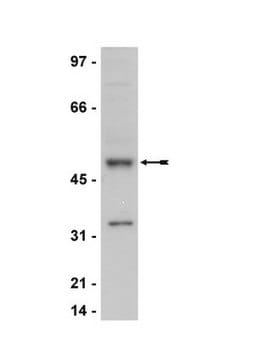
Western Blotting Analysis: 0.1 mg/mL of this antibody detected Cyclin B1 in A431 cell lysate.
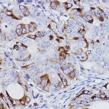
Immunohistochemistry (Paraffin) Analysis: A 1:250 and 1:50 dilutions of this antibody detected Cyclin B1 in Human tonsil and Human bone marrow tissue sections, respectively.
품질
Routinely evaluated by western blot analysis on RIPA lysate from human A431 carcinoma cells.
Western Blot Analysis:
0.5-2 μg/mL of this lot detected cyclin B1 in RIPA lysates from human A431 carcinoma cells.
Western Blot Analysis:
0.5-2 μg/mL of this lot detected cyclin B1 in RIPA lysates from human A431 carcinoma cells.
표적 설명
58 kDa
결합
Replaces: 04-220
물리적 형태
Format: Purified
Protein G purified mouse IgGs in storage buffer containing 0.1 M Tris-glycine, pH 7.4, 0.15 M NaCl, 0.05% sodium azide. Frozen at -20°C.
분석 메모
Control
Positive Antigen Control: Catalog #12-301, non-stimulated A431 cell lysate. Add 2.5µL of 2-mercaptoethanol/100µL of lysate and boil for 5 minutes to reduce the preparation. Load 20µg of reduced lysate per lane for minigels.
Positive Antigen Control: Catalog #12-301, non-stimulated A431 cell lysate. Add 2.5µL of 2-mercaptoethanol/100µL of lysate and boil for 5 minutes to reduce the preparation. Load 20µg of reduced lysate per lane for minigels.
기타 정보
Concentration: Please refer to the Certificate of Analysis for the lot-specific concentration.
법적 정보
UPSTATE is a registered trademark of Merck KGaA, Darmstadt, Germany
Not finding the right product?
Try our 제품 선택기 도구.
시험 성적서(COA)
제품의 로트/배치 번호를 입력하여 시험 성적서(COA)을 검색하십시오. 로트 및 배치 번호는 제품 라벨에 있는 ‘로트’ 또는 ‘배치’라는 용어 뒤에서 찾을 수 있습니다.
Antiproliferation and radiosensitization of caffeic acid phenethyl ester on human medulloblastoma cells.
Lin, Yi-Hsien, et al.
Cancer Chemotherapy and Pharmacology, 57, 525-532 (2006)
Valosin-containing protein is a multi-ubiquitin chain-targeting factor required in ubiquitin-proteasome degradation.
R M Dai, C C Li
Nature Cell Biology null
Matthew S Fuller et al.
Journal of virology, 91(14) (2017-04-28)
Replication of minute virus of mice (MVM) induces a sustained cellular DNA damage response (DDR) which the virus then exploits to prepare the nuclear environment for effective parvovirus takeover. An essential aspect of the MVM-induced DDR is the establishment of
Glyceraldehyde 3-phosphate dehydrogenase is a SET-binding protein and regulates cyclin B-cdk1 activity.
Carujo, S; Estanyol, JM; Ejarque, A; Agell, N; Bachs, O; Pujol, MJ
Oncogene null
Rac1-dependent recruitment of PAK2 to G2 phase centrosomes and their roles in the regulation of mitotic entry.
May, M; Schelle, I; Brakebusch, C; Rottner, K; Genth, H
Cell Cycle null
자사의 과학자팀은 생명 과학, 재료 과학, 화학 합성, 크로마토그래피, 분석 및 기타 많은 영역을 포함한 모든 과학 분야에 경험이 있습니다..
고객지원팀으로 연락바랍니다.


