ABN1720
Anti-Ninein
serum, from rabbit
Synonym(s):
Ninein, Glycogen synthase kinase 3 beta-interacting protein, GSK3B-interacting protein
About This Item
Recommended Products
biological source
rabbit
Quality Level
antibody form
serum
antibody product type
primary antibodies
clone
polyclonal
species reactivity
rat, human, mouse
technique(s)
immunocytochemistry: suitable
immunofluorescence: suitable
immunohistochemistry: suitable
western blot: suitable
NCBI accession no.
UniProt accession no.
shipped in
ambient
target post-translational modification
unmodified
Gene Information
mouse ... Nin(18080)
General description
Specificity
Immunogen
Application
Immunocytochemistry Analysis: A 1:500 dilution from a representative lot detected cytoplasmic and centrosome ninein immunoreactivity in 0.1 Triton X-100 extracted and 4% paraformaldehyde-fixed HEK293T cells (Courtesy of Professor Kensuke Hayashi, Sophia Univesity, Japan).
Immunofluorescence Analysis: A representative lot detected ninein immunoreactivity in the centrosome of proliferative apical progenitor cells (APs) by fluorescent immunohistochemisty staining of 4% paraformaldehyde-fixed cerebral cortex cryosections from E17.5 rat embryos. Ninein immunoreactivity was found downregulated in embryo sections from small eye rats with homozygous rSey2/rSey2 Pax6 mutation (Shinohara, H., et al. (2013). Biol Open. 2(7):739-749).
Immunofluorescence Analysis: A representative lot detected intense ninein immunoreactivity at the ventricular surface by fluorescent immunohistochemisty staining of 4% paraformaldehyde-fixed cerebral cortex cryosections from E13 and E18 mouse embryos. Lower immunoreactivity was also observed in the E18 cortical plate (CP), but was of much weaker intensity in the E13 cortex (Ohama, Y., and Hayashi, K. (2009). Histochem. Cell Biol. 132(5):515-524).
Immunocytochemistry Analysis: A representative lot detected granular ninein immunoreactivity in somatodendritic compartment, but not in the axon by fluorescent immunocytochemistry staining of 4% paraformaldehyde-fixed cultured cortical neurons from E18 mouse embryos. About 60% of ninein granules are associated with microtubules and showed resistance to 0.5% Triton X-100 (Ohama, Y., and Hayashi, K. (2009). Histochem. Cell Biol. 132(5):515-524).
Immunohistochemistry Analysis: A representative lot detected ninein immunoreactivity in the cortical plate (CP), white matter (WM), and the ventricular zone (VZ) of paraffin-embedded embryonic E16.5 mouse brain sections. Ninein immunoreactivity was found downregulated in the CP and WM, but not VZ, of Sip1fl/flNexCre embryos with hippocampal granule cell-targeted Sip1 knockout (Srivatsa, S., et al. (2015). Neuron 85(5):998-1012).
Western Blotting Analysis: A representative lot detected ninein in cortical lysate from postnatal P0, P3, P4 and P7 mouse brain tissue homogenates (Srivatsa, S., et al. (2015). Neuron 85(5):998-1012).
Note: Extracting soluble ninein under microtubule-stabilizing conditions with 0.5% Triton X-100 in the presence of 10 M paclitaxel (Cat. Nos. 580555 & 580556) prior to fixation can be employed to better visualize microtubule- and centrosome-associated ninein immunoreactivity (Ohama, Y., and Hayashi, K. (2009). Histochem. Cell Biol. 132(5):515-524).
Quality
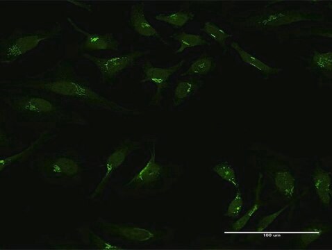
Immunocytochemistry Analysis (ICC): A 1:500 dilution of this antiserum detected ninein in NIH/3T3 cells..
Target description
Other Notes
Not finding the right product?
Try our Product Selector Tool.
Storage Class Code
12 - Non Combustible Liquids
WGK
WGK 1
Flash Point(F)
Not applicable
Flash Point(C)
Not applicable
Certificates of Analysis (COA)
Search for Certificates of Analysis (COA) by entering the products Lot/Batch Number. Lot and Batch Numbers can be found on a product’s label following the words ‘Lot’ or ‘Batch’.
Already Own This Product?
Find documentation for the products that you have recently purchased in the Document Library.
Our team of scientists has experience in all areas of research including Life Science, Material Science, Chemical Synthesis, Chromatography, Analytical and many others.
Contact Technical Service