おすすめの製品
由来生物
rabbit
品質水準
抗体製品の状態
serum
抗体製品タイプ
primary antibodies
クローン
polyclonal
形状
liquid
含みます
≤0.1% sodium azide as preservative
化学種の反応性
porcine, human, rat, mouse
メーカー/製品名
Calbiochem®
保管条件
OK to freeze
avoid repeated freeze/thaw cycles
アイソタイプ
IgG
輸送温度
wet ice
保管温度
−20°C
ターゲットの翻訳後修飾
unmodified
遺伝子情報
human ... UCHL1(7345)
詳細
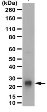
Rabbit polyclonal antibody supplied as undiluted serum. Recognizes the ~26 kDa PGP9.5 protein.
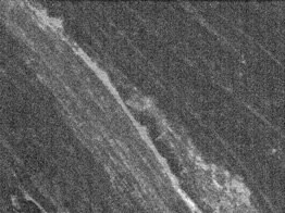
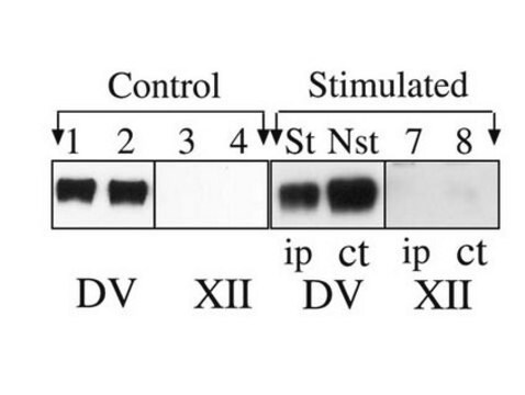
Recognizes the ~26 kDa PGP9.5 protein in mouse dorsal root nerve fibers.
This Anti-PGP9.5 (176-191) Rabbit pAb is validated for use in Immunoblotting, Frozen Sections for the detection of PGP9.5 (176-191).
免疫原
a synthetic peptide (ASSEDTLLKDAAKVCR) corresponding to amino acids 176-191 of PGP9
アプリケーション

Immunoblotting (1:1000)
Frozen Sections (1:2000, fluorescence)
警告
Toxicity: Standard Handling (A)
物理的形状
Undiluted serum.
再構成
Following initial thaw, aliquot and freeze (-20°C).
アナリシスノート
Positive Control
Mouse dorsal root nerve fibers
Mouse dorsal root nerve fibers
その他情報
Caballero, O.L., et al. 2002. Oncogene21, 3003.
Tezel, E., et al. 2000. Clin. Cancer Res.6, 4764.
Tezel, E., et al. 2000. Clin. Cancer Res.6, 4764.
PGP9.5 is also overexpressed in some cancers.
Immunofluorescence protocol
Care should be taken so that the incubation solutions do not evaporate. It is recommended that incubations be performed in a humidified chamber to prevent evaporation. Washing steps should be done in large volumes of phosphate buffered saline (PBS).
1. Warm slides to room temperature.
2. Re-hydrate with PBS for 10-15 min.
3. Remove PBS and incubate in blocking buffer (PBS containing 3% normal donkey serum, 1% bovine serum albumin, 0.3% Triton™ X-100 detergent, and 0.01% NaN3, pH 7.45) for 1 h at room temperature.
4. Dilute primary antibody in blocking buffer. Store diluted antibody at 4°C until use.
5. Remove blocking buffer and add diluted primary antibody.
6. Incubate with primary antibody at 4°C for 18-48 h.
7. Remove primary antibody and wash three times for 10 min with PBS.
8. Dilute secondary antibody in blocking buffer to appropriate dilution (follow manufacture′s recommendation). Store at 4°C until use.
9. Incubate slides in diluted secondary antibody solution for one hour at room temperature.
10. Remove secondary antibody and wash three times for 10 min with PBS.
11. Coverslip slides with appropriate mounting media.
法的情報
CALBIOCHEM is a registered trademark of Merck KGaA, Darmstadt, Germany
Triton is a trademark of The Dow Chemical Company or an affiliated company of Dow
適切な製品が見つかりませんか。
製品選択ツール.をお試しください
保管分類コード
10 - Combustible liquids
WGK
WGK 1
適用法令
試験研究用途を考慮した関連法令を主に挙げております。化学物質以外については、一部の情報のみ提供しています。 製品を安全かつ合法的に使用することは、使用者の義務です。最新情報により修正される場合があります。WEBの反映には時間を要することがあるため、適宜SDSをご参照ください。
Jan Code
NE1013-UL:
NE1013-50UL:
試験成績書(COA)
製品のロット番号・バッチ番号を入力して、試験成績書(COA) を検索できます。ロット番号・バッチ番号は、製品ラベルに「Lot」または「Batch」に続いて記載されています。
J Nakano et al.
Physiological research, 61(6), 643-647 (2012-10-27)
This study was designed to investigate histological changes in skin tissue accompanying immobilization-induced hypersensitivity. Changes in mechanical sensitivity, epidermal thickness, and peripheral nerve profiles in the upper dermis were examined in glabrous skin of rat hind paw after 1, 2
Masato Ogura et al.
PloS one, 16(2), e0246630-e0246630 (2021-02-05)
Interleukin-31 (IL-31) is involved in excessive development of cutaneous sensory nerves in atopic dermatitis (AD), leading to severe pruritus. We previously reported that PQA-18, a prenylated quinolinecarboxylic acid (PQA) derivative, is an immunosuppressant with inhibition of p21-activated kinase 2 (PAK2)
Antonella S Ríos et al.
Development (Cambridge, England), 149(16) (2022-07-30)
The perception of noxious environmental stimuli by nociceptive sensory neurons is an essential mechanism for the prevention of tissue damage. Etv4 is a transcriptional factor expressed in most nociceptors in dorsal root ganglia (DRG) during the embryonic development. However, its
Sandra Boesmueller et al.
Journal of clinical medicine, 8(12) (2019-12-11)
The type II superior labrum anterior to posterior (SLAP) repair is a viable option in young and demanding patients, although a prolonged period of pain after surgery is described in the literature. The reason for this fact remains unknown. Thus
ライフサイエンス、有機合成、材料科学、クロマトグラフィー、分析など、あらゆる分野の研究に経験のあるメンバーがおります。.
製品に関するお問い合わせはこちら(テクニカルサービス)


