詳細
Serine/threonine-protein kinase TAO3 (UniProt: Q9H2K8; also known as EC:2.7.11.1, Cutaneous T-cell lymphoma-associated antigen HD-CL-09, CTCL-associated antigen HD-CL-09, Dendritic cell-derived protein kinase, JNK/SAPK-inhibitory kinase, Jun kinase-inhibitory kinase, JIK, Kinase from chicken homolog A, hKFC-A, Thousand and one amino acid protein 3) is encoded by the TAOK3 (also known as DPK, JIK, KDS, MAP3K18) gene (Gene ID: 51347) in human. TAO kinase 3 is a member of the STE20 subfamily of the STE Ser/Thr protein kinases. It is a serine/threonine-protein kinase that acts as a regulator of the p38/MAPK14 stress-activated MAPK cascade and of the MAPK8/JNK cascade. In response to DNA damage at the G2/M phase, it is phosphorylated at Ser324 by ATM and acts as an activator of the p38/MAPK14 stress-activated MAPK cascade. It is shown to inhibit basal activity of MAPK8/JNK cascade and also diminishes its activation in response epidermal growth factor. TAO kinase 3 is highly expressed in peripheral blood leukocytes, thymus, spleen, kidney, skeletal muscle, heart, and liver.
特異性
Clone 1H9.1 specifically detects Serine/threonine-protein kinase TAO3 (TAOK3) in K562 cells. It targets an epitope of 16 amino acids in the C-terminal region.
免疫原
KLH-conjugated linear peptide corresponding to 16 amino acids from the C-terminus end of human Serine/threonine-protein kinase TAO3 (TAOK3).
アプリケーション
Research Category
細胞シグナル伝達
Detect TAOK3 using this mouse monoclonal Anti-TAOK3 Antibody, clone 1H9.1, Cat. No. MABS1882. It has been tested in Immunohistochemistry (Paraffin) and Western Blotting.
Immunohistochemistry Analysis: A 1:50 dilution from a representative lot detected TAOK3 in human testis, human small intestine, and human pancreas.
Western Blotting Analysis: 0.5 µg/mL from a representative lot detected TAOK3 in 10 µg of Raw264.7 cell lysate.
品質
Evaluated by Western Blotting in K562 cells.
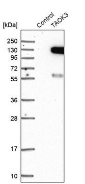
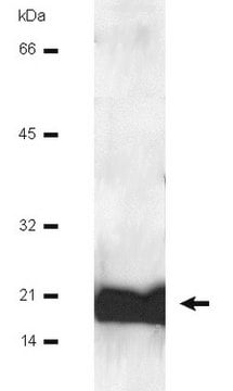
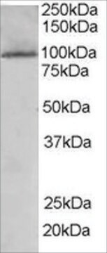
Western Blotting Analysis: 0.5 µg/mL of this antibody detected TAOK3 in 10 µg of K562 cell lysate.
ターゲットの説明
~120 kDa observed, 105.41 kDa calculated. Uncharacterized bands may be observed in some lysate(s).
物理的形状
Protein G purified
Format: Purified
Purified mouse monoclonal antibody IgG2a in buffer containing 0.1 M Tris-Glycine (pH 7.4), 150 mM NaCl with 0.05% sodium azide.
保管および安定性
Stable for 1 year at 2-8°C from date of receipt.
その他情報
Concentration: Please refer to lot specific datasheet.
免責事項
Unless otherwise stated in our catalog or other company documentation accompanying the product(s), our products are intended for research use only and are not to be used for any other purpose, which includes but is not limited to, unauthorized commercial uses, in vitro diagnostic uses, ex vivo or in vivo therapeutic uses or any type of consumption or application to humans or animals.
