おすすめの製品
由来生物
mouse
抗体製品の状態
purified antibody
抗体製品タイプ
primary antibodies
クローン
4G7, monoclonal
化学種の反応性
Ebola virus, virus
包装
antibody small pack of 25 μg
テクニック
ELISA: suitable
immunocytochemistry: suitable
immunoprecipitation (IP): suitable
neutralization: suitable
アイソタイプ
IgG2aκ
NCBIアクセッション番号
UniProtアクセッション番号
ターゲットの翻訳後修飾
unmodified
詳細
Envelope glycoprotein (UniProt: Q05320; also known as GP1,2; GP) is encoded by the GP gene (Gene ID: 911829) in Zaire Ebola virus. Ebola virus (EBOV) is a filovirus that is shown to cause severe viral hemorrhagic fever with high lethality. It has a single stranded, negative-sense RNA genome that encodes seven viral structural proteins including nucleoprotein, virion proteins, and glycoprotein. The glycoprotein is the most important protein involved in pathogenesis. It is a type I transmembrane surface protein shown to be responsible for receptor binding, viral entry, and cellular tropism. The GP gene is reported to undergo transcriptional editing to give rise to several glycosylated proteins, including the structural protein GP1,2, and the secreted non-structural glycoprotein (sGP). The GP is synthesized as a 676-amino acid polyprotein with a signal peptide (aa 1-32). This polyprotein is cleaved by the furin to yield two disulfide linked subunits known as GP1 (aa 33-501) and GP2 (aa 502-676), which together form the GP1,2 heterodimer. The GP1 acts as the receptor-binding subunit and GP2 as the membrane fusion subunit. The GP1 subunit is expressed on the cell surface with the C-terminus oriented toward the aqueous environment, while the N-terminus is bound to GP2 via disulfide bonds. The GP1 subunit contains the heavily glycosylated mucin-like domain (aa 305-485), which is responsible for its cytotoxic function. The glycoprotein contains two coiled coil regions (aa 554-595 and 615-634) that play a role in its oligomerization and fusion activity. Clone 4G7 recognizes glycoprotein lacking a large part of the mucin domain (GP1,2 Mucin333 458). When administered before virus challenge, it provides complete protection. (Ref.: Qiu, X., et al. (2011). Clin. Immunol. 141(2); 218-227).
特異性
Clone 4G7 is a mouse monoclonal antibody that detects envelope glycoprotein of Zaire Ebola virus. It binds to the C-terminal region of GP1.
免疫原
Glycoprotein from Zaire Ebola virus, strain Mayinga, expressed in VSV G. Reverse genetics technique was used to create the recombinant virus VSV G/ZEBOV glycoprotein.
アプリケーション
Research Category
炎症及び免疫
炎症及び免疫
Anti-EBOV GP, clone 4G7, Cat. No. MABF2111, is a mouse monoclonal antibody that detects Envelope glycoprotein in Ebola virus and has been tested for use in ELISA, Immunocytochemistry, Immunoprecipitation, and Neutralizing applications.
Immunocytochemistry Analysis: A representative lot detected EBOV GP in Immunocytochemistry applications (Qiu, X., et. al. (2011). Clin Immunol. 141(2):218-27).
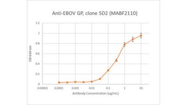
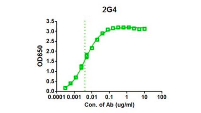
ELISA Analysis: Various dilutions of this antibody detected Envelope glycoprotein in Recombinant Ebola virus Glycoprotein minus the transmembrane region (EBOV rGP TM).
ELISA Analysis: A representative lot detected EBOV GP in ELISA applications (Qiu, X., et. al. (2011). Clin Immunol. 141(2):218-27).
Immunoprecipitation Analysis: A representative lot detected EBOV GP in Immunoprecipitation applications (Qiu, X., et. al. (2011). Clin Immunol. 141(2):218-27).
Neutralizing Analysis: A representative lot provided complete protection when administered prior to virus challenge. (Qiu, X., et. al. (2011). Clin Immunol. 141(2):218-27).
品質
Evaluated by ELISA with recombinant Ebola virus Glycoprotein minus the transmembrane region (EBOV rGP deltaTM).
ELISA Analysis: Various dilutions of this antibody detected EBOV GP in Recombinant Ebola virus Glycoprotein minus the transmembrane region (EBOV rGP deltaTM).
ELISA Analysis: Various dilutions of this antibody detected EBOV GP in Recombinant Ebola virus Glycoprotein minus the transmembrane region (EBOV rGP deltaTM).
ターゲットの説明
74.46 kDa calculated.
物理的形状
Protein G purified
Format: Purified
Purified mouse monoclonal antibody IgG2a in PBS without azide.
保管および安定性
Stable for 1 year at -20°C from date of receipt. Handling Recommendations: Upon receipt and prior to removing the cap, centrifuge the vial and gently mix the solution. Aliquot into microcentrifuge tubes and store at -20°C. Avoid repeated freeze/thaw cycles, which may damage IgG and affect product performance.
その他情報
Concentration: Please refer to lot specific datasheet.
免責事項
Unless otherwise stated in our catalog or other company documentation accompanying the product(s), our products are intended for research use only and are not to be used for any other purpose, which includes but is not limited to, unauthorized commercial uses, in vitro diagnostic uses, ex vivo or in vivo therapeutic uses or any type of consumption or application to humans or animals.
適切な製品が見つかりませんか。
製品選択ツール.をお試しください
試験成績書(COA)
製品のロット番号・バッチ番号を入力して、試験成績書(COA) を検索できます。ロット番号・バッチ番号は、製品ラベルに「Lot」または「Batch」に続いて記載されています。
Viktor Lemmens et al.
NPJ vaccines, 8(1), 99-99 (2023-07-12)
Ebola virus (EBOV) and related filoviruses such as Sudan virus (SUDV) threaten global public health. Effective filovirus vaccines are available only for EBOV, yet restricted to emergency use considering a high reactogenicity and demanding logistics. Here we present YF-EBO, a
ライフサイエンス、有機合成、材料科学、クロマトグラフィー、分析など、あらゆる分野の研究に経験のあるメンバーがおります。.
製品に関するお問い合わせはこちら(テクニカルサービス)