Skip to Content
Merck
All Photos(1)

Documents

05-797R

Sigma-Aldrich

Anti-phospho-Erk1/2 (Thr202/Tyr204, Thr185/Tyr187)Antibody, recombinant clone AW39R, rabbit monoclonal

clone AW39R, Upstate®, from rabbit

Synonym(s):

Erk1, Erk2, p44-MAPK, p42-MAPK, MAPK2, PRKM2, p41mapk, PRKM1, p44, ERK2, P42MAPK, ERK, p42, MAPK1, ERT1, ERT2, Mitogen-activated protein kinase, MAP Kinase 2, MAP Kianse 1, MAPK

Sign Into View Organizational & Contract Pricing


About This Item

UNSPSC Code:
12352203
eCl@ss:
32160702
NACRES:
NA.41

biological source

rabbit

Quality Level

antibody form

purified immunoglobulin

antibody product type

primary antibodies

clone

AW39R, monoclonal

species reactivity

human, mouse, rat

manufacturer/tradename

Upstate®

technique(s)

multiplexing: suitable
western blot: suitable

isotype

IgG

NCBI accession no.

UniProt accession no.

shipped in

dry ice

target post-translational modification

phosphorylation ((pThr202/pTyr204), (pThr185/pTyr187))

Gene Information

human ... MAPK3(5595)
mouse ... Mapk3(26417)
rat ... Mapk3(50689)

General description

Erk (Extracellular signal-Related Kinase) is a family of two, highly homologous proteins denoted as Erk1 (p44, MAPK3) and Erk2 (p42, MAPK1) that both function in the same pathway. The two proteins are often referred to collectively as Erk1/2 or p44/p42 MAP kinase. The Erk pathway is considered the classical, canonical MAPK (Mitogen-Activated Protein Kinase) signaling pathway. It is an evolutionarily conserved pathway that controls and is a critical regulator of the growth and survival through the promotion of cell proliferation and the prevention of apoptosis. Erk is involved in the control of many fundamental cellular processes including cell proliferation, survival, differentiation, apoptosis, motility and metabolism. Erk is activated by growth factor stimulation of receptor tyrosine kinases (RTKs), GPCR, and/or integrin stimulation. This activates the Ras-Raf-MEK-Erk pathway that results in the phosphorylation/activation of Erk1/2 (p44/p42) on the TxY motif (Thr202/Tyr204 and Thr185/Tyr187 for Erk1 & Erk2, respectively).

Specificity

Recognizes Erk 1 & 2 only when dually phosphorylated on its TxY activation motif.

Immunogen

Epitope: pTEpY in the activation loop
Phosphorylated peptide corresponding to amino acids surrounding the pTEpY motif in the activation loop of human phospho-Erk1/2, in which the Thr and Tyr residues are phosphorylated

Application

Anti-phospho-Erk1/2 (Thr202/Tyr204 Antibody, Thr185/Tyr187), recombinant clone AW39R detects level of phospho-Erk1/2 (Thr202/Tyr204 & has been published & validated for use in Mplex & WB.
Research Category
Signaling
Research Sub Category
MAP Kinases

Kinases & Phosphatases

Quality

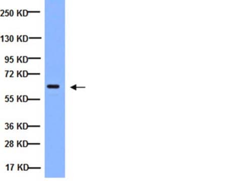
Routinely evaluated by immunoblot on lysate from PDGF-treated NIH/3T3 cells

Target description

Erk1 (p44) (~44 kDa) and Erk2 (p42) (~42 kDa).

Linkage

Replaces: 05-797

Physical form

Format: Purified
Protein A Purified immunoglobulin in 30% glycerol, 0.07M Tris-glycine, pH 7.4, 0.105 M NaCl, 0.035% sodium azide as a preservative.
Protein A purified

Storage and Stability

Stable for 1 year at -20°C from date of receipt. Handling Recommendations: Upon receipt, and prior to removing the cap, centrifuge the vial and gently mix the solution. Aliquot into microcentrifuge tubes and store at -20°C. Avoid repeated freeze/thaw cycles, which may damage IgG and affect product performance.

Analysis Note

Control
PDGF-treated NIH/3T3 cell lysates

Legal Information

UPSTATE is a registered trademark of Merck KGaA, Darmstadt, Germany

Disclaimer

Unless otherwise stated in our catalog or other company documentation accompanying the product(s), our products are intended for research use only and are not to be used for any other purpose, which includes but is not limited to, unauthorized commercial uses, in vitro diagnostic uses, ex vivo or in vivo therapeutic uses or any type of consumption or application to humans or animals.

Not finding the right product?  

Try our Product Selector Tool.

Storage Class Code

12 - Non Combustible Liquids

WGK

WGK 1

Flash Point(F)

Not applicable

Flash Point(C)

Not applicable


Certificates of Analysis (COA)

Search for Certificates of Analysis (COA) by entering the products Lot/Batch Number. Lot and Batch Numbers can be found on a product’s label following the words ‘Lot’ or ‘Batch’.

Already Own This Product?

Find documentation for the products that you have recently purchased in the Document Library.

Visit the Document Library

Wen-Tsan Chang et al.
Molecules (Basel, Switzerland), 22(5) (2017-05-23)
Hepatocellular carcinoma (HCC) is a leading cancer worldwide. Advanced HCCs are usually resistant to anticancer drugs, causing unsatisfactory chemotherapy outcomes. In this study, we showed that a 4-phenoxyphenol derivative, 4-[4-(4-hydroxyphenoxy)phenoxy]phenol (4-HPPP), exerts an inhibitory activity against two HCC cell lines
Crescent R Isham et al.
The Journal of clinical endocrinology and metabolism, 99(6), E936-E943 (2014-03-19)
Vascular endothelial growth factor-targeted kinase inhibitors have emerged as highly promising therapies for radioiodine-refractory metastatic differentiated thyroid cancer. Unfortunately, drug resistance uniformly develops, limiting their therapeutic efficacies and thereby constituting a major clinical problem. To study acquired drug resistance and
Dhia Azzouz et al.
Cell death discovery, 4, 51-51 (2018-05-08)
NETosis is a unique form of neutrophil death that differs from apoptosis and necrosis. However, whether NETosis and apoptosis can occur simultaneously in the same neutrophil is unknown. In this paper, we show that increasing doses of ultraviolet (UV) irradiation
Jiamin Cheng et al.
Oncotarget, 7(15), 20597-20611 (2016-03-12)
Hepatocellular carcinoma (HCC) is the third leading cause of cancer death. This high mortality has been commonly attributed to the presence of residual cancer stem cells (CSCs). Meanwhile, MEK1 signaling is regarded as a key molecular in HCC maintenance and
Chun-Hsu Pan et al.
Aging, 11(19), 8085-8102 (2019-10-05)
The relationship between aging and restenosis are unclear. The purposes of this study were to investigate the possible pathological role and mechanism of aging on formation of restenosis. Our data indicated that cell proliferation and migration of the oxidative stress-induced

Our team of scientists has experience in all areas of research including Life Science, Material Science, Chemical Synthesis, Chromatography, Analytical and many others.

Contact Technical Service