推荐产品
生物源
rabbit
共軛
unconjugated
抗體表格
affinity isolated antibody
抗體產品種類
primary antibodies
無性繁殖
polyclonal
產品線
Prestige Antibodies® Powered by Atlas Antibodies
形狀
buffered aqueous glycerol solution
物種活性
human
加強驗證
orthogonal RNAseq
recombinant expression
Learn more about Antibody Enhanced Validation
技術
immunofluorescence: 0.25-2 μg/mL
immunohistochemistry: 1:50-1:200
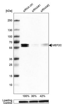
western blot: 0.04-0.4 μg/mL
免疫原序列
PPNFEMPVSIPVSSHNSLVYSNPVSSLGNPNLLPLAHPSLQRNSMSPGVTHRPPSAGNTGGLMGGDLTSGAGTSAGNGYGNPRNSPGLLVSPGNLNKNMQAKSPPPMNLGMNNRKPDLRVLIPPGSKNTMPSVNQRINN
UniProt登錄號
運輸包裝
wet ice
儲存溫度
−20°C
目標翻譯後修改
unmodified
基因資訊
human ... MEF2C(4208)
一般說明
MEF2C (myocyte enhancer factor 2C) belongs to Myocyte enhancer factor 2 (MEF2) protein family, which in turn belongs to a family of transcriptional regulators called MADS (MCMI, agamous, deficiens, serum response factor)-box. Four different forms of MEF2 protein are found in vertebrates, namely, MEF2A, MEF2B, MEF2C, and MEF2D. MEF2C is predominantly expressed in brain and skeletal muscle. It shares the common DNA-binding and dimerization domain present at the N-terminal, with other MEF2 proteins. Alternative splicing produces two MEF2C variants, which lack α exon. These MEF2Cα- are ubiquitously expressed, but at a lower level than MEF2Cα+ variants. MEF2Cα- variants are also less expressed in other tissues, as opposed to brain and heart. Alternative splicing also produces either MEF2Cγ+ or MEF2Cγ- isoforms. MEF2Cγ- is the major isoform expressed in differentiating myocytes and adult tissues. MEF2C is mapped to chromosome 5q14.3.
免疫原
Myocyte-specific enhancer factor 2C recombinant protein epitope signature tag (PrEST)
應用
Anti-MEF2C antibody is suitable for chromatin immunoprecipitation (ChIP).
Anti-MEF2C antibody produced in rabbit, a Prestige Antibody, is developed and validated by the Human Protein Atlas (HPA) project . Each antibody is tested by immunohistochemistry against hundreds of normal and disease tissues. These images can be viewed on the Human Protein Atlas (HPA) site by clicking on the Image Gallery link. The antibodies are also tested using immunofluorescence and western blotting. To view these protocols and other useful information about Prestige Antibodies and the HPA, visit sigma.com/prestige.
Anti-MEF2C antibody produced in rabbit, a Prestige Antibody, is developed and validated by the Human Protein Atlas (HPA) project . Each antibody is tested by immunohistochemistry against hundreds of normal and disease tissues. These images can be viewed on the Human Protein Atlas (HPA) site by clicking on the Image Gallery link. The antibodies are also tested using immunofluorescence and western blotting. To view these protocols and other useful information about Prestige Antibodies and the HPA, visit sigma.com/prestige.
Applications in which this antibody has been used successfully, and the associated peer-reviewed papers, are given below.
Immunohistochemistry (1 paper)
Immunohistochemistry (1 paper)
生化/生理作用
MEF2C (myocyte enhancer factor 2C) belongs to the family of transcription factors, which regulate gene expression in myocytes, neurons and lymphocytes. BMK1 phosphorylates and activates MEF2C. Serum also induces BMK1-induced phosphorylation of MEF2C, and thus, MEF2C plays a role in serum-dependent early gene expression via BMK1 pathway. Lipopolysaccharides produced during microbial infection activate this protein via phosphorylation by p38. This induces c-jun transcription, which plays a role in inflammation. MEF2C plays a part in neuronal differentiation, as it is expressed in cortical plate, during early development. It is also expressed in neurons having a preference to mature cerebrocortex layers II, IV and VI. MEF2C contributes to early pathogenesis of Parkinson′s disease, as the disruption of MEF2C- PGC1α pathway, leads to neuronal apoptosis due to mitochondrial dysfunction. It is a part of Wnt pathway, and hence plays a role in control of bone mass and turnover.
特點和優勢
Prestige Antibodies® are highly characterized and extensively validated antibodies with the added benefit of all available characterization data for each target being accessible via the Human Protein Atlas portal linked just below the product name at the top of this page. The uniqueness and low cross-reactivity of the Prestige Antibodies® to other proteins are due to a thorough selection of antigen regions, affinity purification, and stringent selection. Prestige antigen controls are available for every corresponding Prestige Antibody and can be found in the linkage section.
Every Prestige Antibody is tested in the following ways:
Every Prestige Antibody is tested in the following ways:
- IHC tissue array of 44 normal human tissues and 20 of the most common cancer type tissues.
- Protein array of 364 human recombinant protein fragments.
聯結
Corresponding Antigen APREST79903
外觀
Solution in phosphate-buffered saline, pH 7.2, containing 40% glycerol and 0.02% sodium azide
法律資訊
Prestige Antibodies is a registered trademark of Merck KGaA, Darmstadt, Germany
免責聲明
Unless otherwise stated in our catalog or other company documentation accompanying the product(s), our products are intended for research use only and are not to be used for any other purpose, which includes but is not limited to, unauthorized commercial uses, in vitro diagnostic uses, ex vivo or in vivo therapeutic uses or any type of consumption or application to humans or animals.
Not finding the right product?
Try our 产品选型工具.
儲存類別代碼
10 - Combustible liquids
水污染物質分類(WGK)
WGK 1
閃點(°F)
Not applicable
閃點(°C)
Not applicable
個人防護裝備
Eyeshields, Gloves, multi-purpose combination respirator cartridge (US)
Scott D Ryan et al.
Cell, 155(6), 1351-1364 (2013-12-03)
Parkinson's disease (PD) is characterized by loss of A9 dopaminergic (DA) neurons in the substantia nigra pars compacta (SNpc). An association has been reported between PD and exposure to mitochondrial toxins, including environmental pesticides paraquat, maneb, and rotenone. Here, using
Dana Trompet et al.
JCI insight, 9(6) (2024-03-22)
Recently, skeletal stem cells were shown to be present in the epiphyseal growth plate (epiphyseal skeletal stem cells, epSSCs), but their function in connection with linear bone growth remains unknown. Here, we explore the possibility that modulating the number of
Christopher D Clark et al.
Mechanisms of development, 162, 103615-103615 (2020-05-26)
The cardiac homeobox transcription factor Nkx2-5 is a major determinant of cardiac identity and cardiac morphogenesis. Nkx2-5 operates as part of a complex and mutually reinforcing network of early transcription factors of the homeobox, GATA zinc finger and MADS domain
J Han et al.
Nature, 386(6622), 296-299 (1997-03-20)
For cells of the innate immune system to mount a host defence response to infection, they must recognize products of microbial pathogens such as lipopolysaccharide (LPS), the endotoxin secreted by Gram-negative bacteria. These cellular responses require intracellular signalling pathways, such
Kewei Ma et al.
Molecular and cellular biology, 25(9), 3575-3582 (2005-04-16)
Myocyte enhancer factor 2 (MEF2) family proteins are key transcription factors controlling gene expression in myocytes, lymphocytes, and neurons. MEF2 proteins are known to be regulated by phosphorylation. We now provide evidence showing that MEF2C is acetylated by p300 both
我们的科学家团队拥有各种研究领域经验,包括生命科学、材料科学、化学合成、色谱、分析及许多其他领域.
联系技术服务部门


