
推荐产品


生物来源
mouse
质量水平
偶联物
unconjugated
抗体形式
purified antibody
抗体产品类型
primary antibodies
克隆
CSTEM29, monoclonal
分子量
calculated mol wt 91.42 kDa
observed mol wt ~80 kDa
纯化方式
using protein G
种属反应性
human
包装
antibody small pack of 100 μg
技术
flow cytometry: suitable
western blot: suitable
同位素/亚型
IgG2aκ
表位序列
Extracellular domain
Protein ID登记号
UniProt登记号
运输
2-8°C
靶向翻译后修饰
unmodified
基因信息
human ... CHD3(1001)
一般描述
Cadherin-3 (UniProt: P22223; also known as Placental cadherin, P-cadherin) is encoded by the CHD3 (also known as CDHP) gene (Gene ID: 1001) in human. Cadherins are calcium-dependent cell adhesion proteins that preferentially interact with themselves in a homophilic manner in connecting cells. Cadherins are calcium-dependent cell adhesion proteins that preferentially interact with themselves in a homophilic manner in connecting cells. P-cadherin is a class I cadherin that is expressed in myoepithelial cells and is also expressed during late pregnancy and lactation, when luminal epithelial cells secrete high levels of a soluble fragment of P-cadherin in human milk. It is a single pass type I membrane glycoprotein that is synthesized with a signal peptide (aa 1-24) and a propeptide (aa 25-107 that are subsequently cleaved off to generate the mature form that contains an extracellular domain (aa 108-654), a transmembrane domain (aa 655-677), and a cytoplasmic domain (aa 678-829). It has 5 cadherin domains. Three calcium ions are usually bound at the interface of each cadherin domain and rigidify the connections and thereby imparting a strong curvature to the full-length ectodomain. P-cadherin dysfunction is strongly associated with tumorigenesis and confers the malignant phenotype in cancer cells. Its overexpression is strongly associated with poor prognosis in several solid tumors. Its high expression is reported to be predictive of shorter overall survival in glioblastoma patients. It is also detected in plasma membrane during progression of pancreatic intraepithelial ductal adenocarcinoma. (Ref.: Martins, EP., et al. (2021). Mol. Oncol. doi: 10.1002/1878-0261; Yu, W., et al. (2019). Front. Oncol. 9; 989; Siret, C., et al. (2018). Br. J. Cancer. 118; 546-557).
特异性
Clone CSTEM3 is a mouse monoclonal antibody that detects P-Cadherin (Cadherin 3). It targets an epitope within the extracellular domain.
免疫原
His-tagged recombinant protein fragment corresponding to the extracellular domain of human P-Cadherin (Cadherin 3).
应用
Quality Control Testing
Evaluated by Western Blotting with recombinant human P-Cadherin.
Western Blotting Analysis: A 1:1,000 dilution of this antibody detected recombinant Human P-Cadherin.
Tested Applications
ELISA Analysis: A representative lot detected P-Cadherin in ELISA applications (O Brien, C.M., et al. (2017). Stem Cells. 35(3); 626-640).
Flow Cytometry Analysis: A representative lot detected P-Cadherin in Flow Cytometry applications (O Brien, C.M., et al. (2017). Stem Cells. 35(3); 626-640).
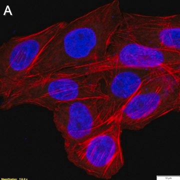
Immunocytochemistry Analysis: A representative lot detected P-Cadherin in Immunocytochemistry applications (O Brien, C.M., et al. (2017). Stem Cells. 35(3); 626-640).
Note: Actual optimal working dilutions must be determined by end user as specimens, and experimental conditions may vary with the end user
Evaluated by Western Blotting with recombinant human P-Cadherin.
Western Blotting Analysis: A 1:1,000 dilution of this antibody detected recombinant Human P-Cadherin.
Tested Applications
ELISA Analysis: A representative lot detected P-Cadherin in ELISA applications (O Brien, C.M., et al. (2017). Stem Cells. 35(3); 626-640).
Flow Cytometry Analysis: A representative lot detected P-Cadherin in Flow Cytometry applications (O Brien, C.M., et al. (2017). Stem Cells. 35(3); 626-640).
Immunocytochemistry Analysis: A representative lot detected P-Cadherin in Immunocytochemistry applications (O Brien, C.M., et al. (2017). Stem Cells. 35(3); 626-640).
Note: Actual optimal working dilutions must be determined by end user as specimens, and experimental conditions may vary with the end user
Anti-P-Cadherin, clone CSTEM29, Cat. No. MABT1346, is a mouse monoclonal antibody that detects P-Cadherin and is tested for use in ELISA, Flow Cytometry, Immunocytochemistry, and Western Blotting.
外形
Purified mouse monoclonal antibody IgG2a in buffer containing 0.1 M Tris-Glycine (pH 7.4), 150 mM NaCl with 0.05% sodium azide.
储存及稳定性
Recommended storage: +2°C to +8°C.
其他说明
Concentration: Please refer to the Certificate of Analysis for the lot-specific concentration.
免责声明
Unless otherwise stated in our catalog or other company documentation accompanying the product(s), our products are intended for research use only and are not to be used for any other purpose, which includes but is not limited to, unauthorized commercial uses, in vitro diagnostic uses, ex vivo or in vivo therapeutic uses or any type of consumption or application to humans or animals.
未找到合适的产品?
试试我们的产品选型工具.
储存分类代码
12 - Non Combustible Liquids
WGK
WGK 1
闪点(°F)
Not applicable
闪点(°C)
Not applicable
我们的科学家团队拥有各种研究领域经验,包括生命科学、材料科学、化学合成、色谱、分析及许多其他领域.
联系技术服务部门