AB2226
Anti-MDGA1 Antibody
from rabbit, purified by affinity chromatography
别名:
MAM domain-containing glycosylphosphatidylinositol anchor protein 1, GPI and MAM protein, GPIM, Glycosylphosphatidylinositol-MAM, MAM domain-containing protein 3
登录查看公司和协议定价
所有图片(1)
About This Item
分類程式碼代碼:
12352203
eCl@ss:
32160702
NACRES:
NA.41
推荐产品
生物源
rabbit
品質等級
抗體表格
affinity isolated antibody
抗體產品種類
primary antibodies
無性繁殖
polyclonal
純化經由
affinity chromatography
物種活性
mouse, rat
物種活性(以同源性預測)
human (based on 100% sequence homology), bovine (based on 100% sequence homology)
技術
western blot: suitable
NCBI登錄號
UniProt登錄號
運輸包裝
wet ice
目標翻譯後修改
unmodified
基因資訊
human ... MDGA1(266727)
一般說明
Studies report that MDGA1 is a layer-specific marker and an area-specific marker, being expressed in layers 2/3 throughout the neocortex, but within the primary somatosensory area (S1), MDGA1 is also uniquely expressed in layers 4 and 6a. Comparisons with other markers, including cadherins, serotonin, cytochrome oxidase, RORß, and COUP-TF1, reveal unique features of patterned expression of MDGA1 within cortex and S1 barrels. Furthermore, during early stages of development, MDGA1 is expressed by Reelin- and Tbr1-positive Cajal–Retzius neurons that originate from multiple sources outside of neocortex and emigrate into it. At even earlier stages, MDGA1 is expressed by the earliest diencephalic and mesencephalic neurons, which appear to migrate from a MDGA1-positive domain of progenitors in the diencephalon and form a "preplate." These findings show that MDGA1 is a unique marker for studies of cortical lamination and area patterning and together with recent reports suggest that MDGA1 has critical functions in forebrain/midbrain development.
特異性
This antibody recognizes the MAM domain of MDGA1.
免疫原
Epitope: MAM domain
KLH-conjugated linear peptide corresponding to the MAM domain of MDGA1.
應用
Anti-MDGA1 Antibody detects level of MDGA1 & has been published & validated for use in Western Blotting.
Research Category
Neuroscience
Neuroscience
Research Sub Category
Developmental Neuroscience
Developmental Neuroscience
品質
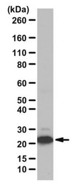
Evaluated by Western Blot in untreated and PNGase F treated mouse P7 brain tissue lysate.
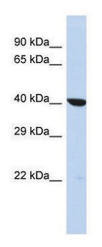
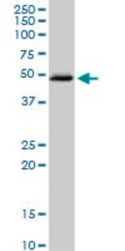
Western Blot Analysis: 0.5 µg/mL of this antibody detected MDGA1 in 5 µg of untreated and PNGase F treated mouse P7 brain tissue lysate. This antibody recognizes glycosolated (>160 kDa) (lane 1) and deglycosolated MDGA1 (lane 2).
Western Blot Analysis: 0.5 µg/mL of this antibody detected MDGA1 in 5 µg of untreated and PNGase F treated mouse P7 brain tissue lysate. This antibody recognizes glycosolated (>160 kDa) (lane 1) and deglycosolated MDGA1 (lane 2).
標靶描述
~130kDa observed. UniProt describes 2 isoforms produced by alternative splicing at ~106 kDa (Isoform 1), ~107 kDa (Isoform 2). An uncharacterized band may be observed at ~48 kDa in some tissue lysates.
外觀
Affinity purified
Purified rabbit polyclonal in buffer containing 0.1 M Tris-Glycine (pH 7.4), 150 mM NaCl with 0.05% sodium azide.
儲存和穩定性
Stable for 1 year at 2-8°C from date of receipt.
分析報告
Control
Untreated and PNGase F treated mouse P7 brain tissue lysate.
Untreated and PNGase F treated mouse P7 brain tissue lysate.
其他說明
Concentration: Please refer to the Certificate of Analysis for the lot-specific concentration.
免責聲明
Unless otherwise stated in our catalog or other company documentation accompanying the product(s), our products are intended for research use only and are not to be used for any other purpose, which includes but is not limited to, unauthorized commercial uses, in vitro diagnostic uses, ex vivo or in vivo therapeutic uses or any type of consumption or application to humans or animals.
Not finding the right product?
Try our 产品选型工具.
儲存類別代碼
12 - Non Combustible Liquids
水污染物質分類(WGK)
WGK 1
閃點(°F)
Not applicable
閃點(°C)
Not applicable
我们的科学家团队拥有各种研究领域经验,包括生命科学、材料科学、化学合成、色谱、分析及许多其他领域.
联系技术服务部门
