推荐产品
生物源
rabbit
品質等級
抗體表格
serum
抗體產品種類
primary antibodies
無性繁殖
polyclonal
物種活性
human, chicken, hamster, rat, mouse
製造商/商標名
Chemicon®
技術
ELISA: suitable
immunocytochemistry: suitable
immunofluorescence: suitable
immunohistochemistry: suitable
immunoprecipitation (IP): suitable
radioimmunoassay: suitable
western blot: suitable
NCBI登錄號
UniProt登錄號
運輸包裝
dry ice
目標翻譯後修改
unmodified
基因資訊
human ... ITGA5(3678)
一般說明
整联蛋白是介导细胞-细胞和细胞外基质粘连的二聚体跨膜蛋白家族。整联蛋白转导的信号在许多生物学过程中起作用,包括细胞生长,分化,迁移和凋亡。整联蛋白家族由至少15个α和8个β亚基组成,其可以在细胞表面上形成超过20种不同的α-β非共价结合的二聚体组合。全部α亚基彼此之间均具有某些同源性,β亚基也是如此。两个亚基都有助于配体的结合。整联蛋白α亚基在N-末端区域包含七个弱序列重复,这可能在配体结合中很重要,并且已预计可以协同折叠成具有七个β-折叠的单个β-螺旋桨域。α-3亚基(CD49c)高度集中在上皮细胞中,在那里它强烈粘附于层粘连蛋白5,层粘连蛋白5诱导的快速粘连可以被针对α-3整联蛋白亚基的抗体阻断。α-3亚基存在于两个不同的剪接变体中,分别表示为“A”和“B”。由这种差异剪接引起的唯一差异是细胞质结构域的总变化,而细胞外结构域保持不变。缺乏该亚基的敲除小鼠显示出产前致死性和肾脏异常。
特異性
与其他物种的反应性尚未确定。
抗体识别整合蛋白α5。通过免疫沉淀从35S甲硫氨酸标记的内皮细胞的去污剂提取物中确定了特异性。与α1、α2、α3、α4、α6 或αV无交叉反应。该抗体可识别蛋白质的胞质结构域上的表位,因此是在细胞内。
免疫原
来源于人α5整联蛋白亚单位C端序列(胞质域)的合成肽。
應用
抗整联蛋白α5抗体,C端,整联蛋白α5细胞内检测水平&已发表,& 经验证可用于ELISA,IC,IF,IH,IP,RIA & WB。
蛋白质印迹分析:
使用先前批次的1:1000稀释液。
ELISA:
先前批次的1:500-1:2000稀释液用于ELISA。
免疫沉淀:
对于5x10⁶个细胞,建议使用5 μL抗体。
免疫组织化学:
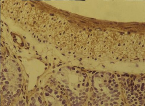
先前批次的1:1000稀释液用于组织染色;建议仅用于丙酮固定的组织。
免疫细胞化学:
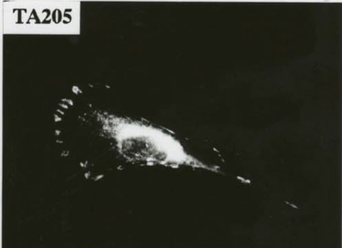
先前批次的1:1000稀释液用于免疫细胞化学。
最佳工作稀释度必须由最终用户确定。
使用先前批次的1:1000稀释液。
ELISA:
先前批次的1:500-1:2000稀释液用于ELISA。
免疫沉淀:
对于5x10⁶个细胞,建议使用5 μL抗体。
免疫组织化学:
先前批次的1:1000稀释液用于组织染色;建议仅用于丙酮固定的组织。
免疫细胞化学:
先前批次的1:1000稀释液用于免疫细胞化学。
最佳工作稀释度必须由最终用户确定。
品質
已通过蛋白质印迹对HUVEC裂解液进行了常规评估。
蛋白质印迹分析:该批次的1:1000稀释液在10 μg HUVEC裂解液中检测到整合蛋白α5。
蛋白质印迹分析:该批次的1:1000稀释液在10 μg HUVEC裂解液中检测到整合蛋白α5。
標靶描述
114kda
外觀
兔多克隆抗血清溶于含0.05%叠氮化钠的缓冲液中。
分析報告
对照
小鼠3T3皮肤成纤维细胞(基底膜)。
小鼠3T3皮肤成纤维细胞(基底膜)。
其他說明
浓度:请参考批次特异性浓缩物的检验报告。
法律資訊
CHEMICON is a registered trademark of Merck KGaA, Darmstadt, Germany
Not finding the right product?
Try our 产品选型工具.
儲存類別代碼
12 - Non Combustible Liquids
水污染物質分類(WGK)
WGK 1
閃點(°F)
Not applicable
閃點(°C)
Not applicable
Mary Ann Stepp et al.
Molecular carcinogenesis, 49(4), 363-373 (2010-01-19)
Syndecan-1 (sdc-1) is a cell surface proteoglycan that mediates the interaction of cells with their matrix, influencing attachment, migration, and response to growth factors. In keratinocytes, loss of sdc-1 delays wound healing, reduces migration, and increases Transforming growth factor beta
Vincent F Fiore et al.
The Journal of cell biology, 211(1), 173-190 (2015-10-16)
Progressive fibrosis is characterized by excessive deposition of extracellular matrix (ECM), resulting in gross alterations in tissue mechanics. Changes in tissue mechanics can further augment scar deposition through fibroblast mechanotransduction. In idiopathic pulmonary fibrosis, a fatal form of progressive lung
Kazuko Goto et al.
Stem cells translational medicine, 5(2), 218-226 (2015-12-25)
When injected directly into ischemic tissue in patients with peripheral artery disease, the reparative capacity of endothelial progenitor cells (EPCs) appears to be limited by their poor survival. We, therefore, attempted to improve the survival of transplanted EPCs through intravenous
Andoria Tjondrokoesoemo et al.
The Journal of biological chemistry, 291(19), 9920-9928 (2016-03-12)
Duchenne muscular dystrophy (DMD) is an X-linked recessive disease caused by mutations in the gene encoding dystrophin. Loss of dystrophin protein compromises the stability of the sarcolemma membrane surrounding each muscle cell fiber, leading to membrane ruptures and leakiness that
Jamie L Marshall et al.
Human molecular genetics, 24(7), 2011-2022 (2014-12-17)
Duchenne muscular dystrophy (DMD) is caused by mutations in the dystrophin gene that result in loss of the dystrophin-glycoprotein complex, a laminin receptor that connects the myofiber to its surrounding extracellular matrix. Utrophin, a dystrophin ortholog that is normally localized
我们的科学家团队拥有各种研究领域经验,包括生命科学、材料科学、化学合成、色谱、分析及许多其他领域.
联系技术服务部门


