Saltar al contenido
Merck
Todas las fotos(1)

Key Documents

06-1013

Sigma-Aldrich

Anti-NET5 Antibody

from rabbit, purified by affinity chromatography

Sinónimos:

transmembrane protein 201, Spindle-associated membrane protein 1

Iniciar sesiónpara Ver la Fijación de precios por contrato y de la organización


About This Item

UNSPSC Code:
12352203
eCl@ss:
32160702
NACRES:
NA.41

biological source

rabbit

Quality Level

antibody form

affinity isolated antibody

antibody product type

primary antibodies

clone

polyclonal

purified by

affinity chromatography

species reactivity

rat, human, mouse

species reactivity (predicted by homology)

orangutan

technique(s)

immunocytochemistry: suitable
western blot: suitable

NCBI accession no.

UniProt accession no.

shipped in

wet ice

target post-translational modification

unmodified

Gene Information

General description

Transmembrane protein 201, also known as NET5, is a transmembrane protein of the nuclear envelope that is homologous to spindle-associated membrane protein 1 (SAMP1) in humans. NET5 is thought to be expressed on the inner nuclear membrane during interphase and move to the polar regions of the mitotic spindle during mitosis.

Immunogen

KLH-conjugated linear peptide corresponding to human NET5.

Application

Research Category
Epigenetics & Nuclear Function
Research Sub Category
Cell Cycle, DNA Replication & Repair
Use Anti-NET5 Antibody (Rabbit Polyclonal Antibody) validated in WB, ICC to detect NET5 also known as transmembrane protein 201.
Western Blot Analysis: 1 µg/mL from a representative lot detected NET5 on 10 µg of rat heart tissue lysate.

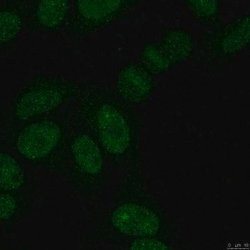
Immunocytochemistry Analysis: A representative lot was used by an independent laboratory that detected NET5 in U2OS cells expressing various NET-GFP fusions. (Wilkie, G., et al. (2011) Mol. Cell Proteomics).

Quality

Evaluated by Western Blot in human heart tissue lysate.

Western Blot Analysis: 1 µg/mL of this antibody detected NET5 on 10 µg of human heart tissue lysate.

Target description

~72 and/or 43 kDa observed depending on lysate used. Isoform1: 72 kDa and Isoform 2: 43 kDa Uncharacterized band at ~28 kDa may be observed in some lysates.

Physical form

Affinity purified
Purified rabbit polyclonal containing 0.1 M Tris-Glycine (pH 7.4), 150 mM NaCl with 0.05% sodium azide.

Storage and Stability

Stable for 1 year at 2-8°C from date of receipt.

Analysis Note

Control
Human heart tissue lysate

Other Notes

Concentration: Please refer to the Certificate of Analysis for the lot-specific concentration.

Disclaimer

Unless otherwise stated in our catalog or other company documentation accompanying the product(s), our products are intended for research use only and are not to be used for any other purpose, which includes but is not limited to, unauthorized commercial uses, in vitro diagnostic uses, ex vivo or in vivo therapeutic uses or any type of consumption or application to humans or animals.

¿No encuentra el producto adecuado?  

Pruebe nuestro Herramienta de selección de productos.

Storage Class

12 - Non Combustible Liquids

wgk_germany

WGK 1

flash_point_f

Not applicable

flash_point_c

Not applicable


Certificados de análisis (COA)

Busque Certificados de análisis (COA) introduciendo el número de lote del producto. Los números de lote se encuentran en la etiqueta del producto después de las palabras «Lot» o «Batch»

¿Ya tiene este producto?

Encuentre la documentación para los productos que ha comprado recientemente en la Biblioteca de documentos.

Visite la Librería de documentos

Anne T Bertrand et al.
Cells, 9(4) (2020-04-05)
LMNA encodes for Lamin A/C, type V intermediate filaments that polymerize under the inner nuclear membrane to form the nuclear lamina. A small fraction of Lamin A/C, less polymerized, is also found in the nucleoplasm. Lamin A/C functions include roles
Nikolaj Zuleger et al.
Genome biology, 14(2), R14-R14 (2013-02-19)
Different cell types have distinctive patterns of chromosome positioning in the nucleus. Although ectopic affinity-tethering of specific loci can be used to relocate chromosomes to the nuclear periphery, endogenous nuclear envelope proteins that control such a mechanism in mammalian cells
Phu Le Thanh et al.
Neuromuscular disorders : NMD, 27(4), 338-351 (2017-02-19)
Reports of aberrant distribution for some nuclear envelope proteins in cells expressing a few Emery-Dreifuss muscular dystrophy mutations raised the possibility that such protein redistribution could underlie pathology and/or be diagnostic. However, this disorder is linked to 8 different genes
Nadia Korfali et al.
Nucleus (Austin, Tex.), 3(6), 552-564 (2012-09-20)
One hypothesis to explain how mutations in the same nuclear envelope proteins yield pathologies focused in distinct tissues is that as yet unidentified tissue-specific partners mediate the disease pathologies. The nuclear envelope proteome was recently determined from leukocytes and muscle.
Gavin S Wilkie et al.
Molecular & cellular proteomics : MCP, 10(1), M110-M110 (2010-09-30)
Nuclear envelopes from liver and a neuroblastoma cell line have previously been analyzed by proteomics; however, most diseases associated with the nuclear envelope affect muscle. To determine whether muscle has unique nuclear envelope proteins, rat skeletal muscle nuclear envelopes were

Nuestro equipo de científicos tiene experiencia en todas las áreas de investigación: Ciencias de la vida, Ciencia de los materiales, Síntesis química, Cromatografía, Analítica y muchas otras.

Póngase en contacto con el Servicio técnico