Accéder au contenu
Merck
Toutes les photos(3)

Key Documents

B0529

Sigma-Aldrich

Anti-Mouse IgG (Fab specific)–Biotin antibody produced in goat

affinity isolated antibody, buffered aqueous solution

Synonyme(s) :

Goat anti-Mouse IgG

Se connecterpour consulter vos tarifs contractuels et ceux de votre entreprise/organisme


About This Item

Numéro MDL:
Code UNSPSC :
12352203
Nomenclature NACRES :
NA.46

Source biologique

goat

Niveau de qualité

Conjugué

biotin conjugate

Forme d'anticorps

affinity isolated antibody

Type de produit anticorps

secondary antibodies

Clone

polyclonal

Forme

buffered aqueous solution

Espèces réactives

mouse

Technique(s)

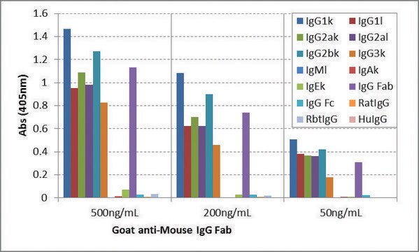
direct ELISA: 1:200,000
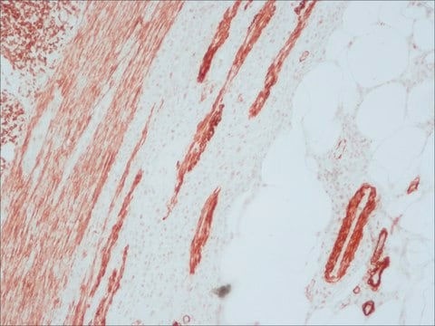
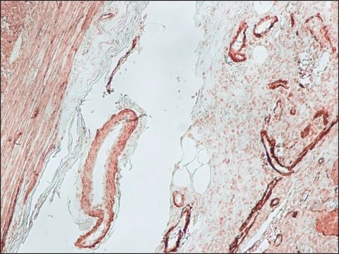
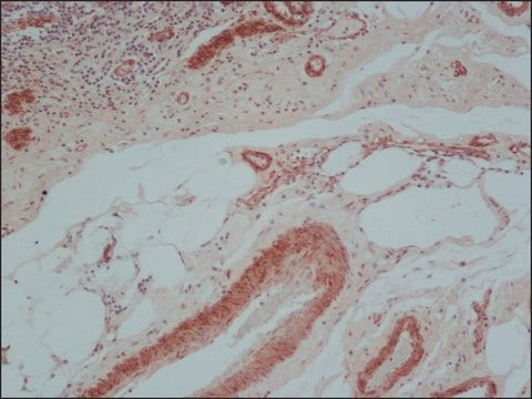
immunohistochemistry (formalin-fixed, paraffin-embedded sections): 1:300
western blot: 1:300,000-1:600,000 using using Hela cells extract

Température de stockage

−20°C

Modification post-traductionnelle de la cible

unmodified

Vous recherchez des produits similaires ? Visite Guide de comparaison des produits

Description générale

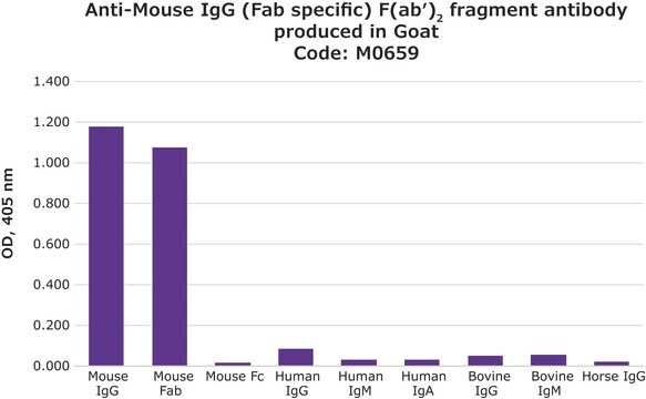
Binds all mouse Igs.
Immunoglobulin G (IgG) belongs to the immunoglobulin family and is a widely expressed serum antibody. Each immunoglobulin has two heavy chains and two light chains connected by a disulfide bond. It is a glycoprotein. IgG is a major class of immunoglobulin. Mouse consists of five immunoglobulin classes- IgM, IgG, IgA, IgD and IgE. Mouse IgG is further divided into five classes- IgG1, IgG2a, IgG2b and IgG3.

Application

Anti-Mouse IgG (Fab specific)-Biotin antibody produced in goat has been used in immunohistochemistry and immunofluorescence studies.
Anti-Mouse IgG (Fab specific)-Biotin antibody produced in goat is suitable for direct ELISA at a dilution of 1:200000 and for immunohistochemistry at a dilution of 1:300. The antibody was used to stain paraffin-embedded, rat skin, duodenum, esophagus and spleen sections at a dilution of 1:200. For immunofluorescence of Human Gingival Fibroblasts, a dilution of 1:200 was used. For immunoblotting experiments using mouse adipose tissue, antibody dilution of 1:30000 was used.

Actions biochimiques/physiologiques

IgG antibody subtype is the most abundant of serum immunoglobulins of the immune system. It is secreted by B cells and is found in blood and extracellular fluids and provides protection from infections caused by bacteria, fungi and viruses. Maternal IgG is transferred to fetus through the placenta that is vital for immune defense of the neonate against infections.
Immunoglobulin G (IgG) participates in hypersensitivity type II and type III reactions. IgG helps in opsonization, complement fixation and antibody dependent cell mediated cytotoxicity.

Autres remarques

Antibody adsorbed with human IgG and rat serum proteins.

Forme physique

Solution in 0.01 M phosphate buffered saline, pH 7.4, containing 15 mM sodium azide.

Notes préparatoires

Adsorbed to reduce background staining with human or rat samples.

Clause de non-responsabilité

Unless otherwise stated in our catalog or other company documentation accompanying the product(s), our products are intended for research use only and are not to be used for any other purpose, which includes but is not limited to, unauthorized commercial uses, in vitro diagnostic uses, ex vivo or in vivo therapeutic uses or any type of consumption or application to humans or animals.

Vous ne trouvez pas le bon produit ?  

Essayez notre Outil de sélection de produits.

Code de la classe de stockage

10 - Combustible liquids

Classe de danger pour l'eau (WGK)

nwg

Point d'éclair (°F)

Not applicable

Point d'éclair (°C)

Not applicable


Certificats d'analyse (COA)

Recherchez un Certificats d'analyse (COA) en saisissant le numéro de lot du produit. Les numéros de lot figurent sur l'étiquette du produit après les mots "Lot" ou "Batch".

Déjà en possession de ce produit ?

Retrouvez la documentation relative aux produits que vous avez récemment achetés dans la Bibliothèque de documents.

Consulter la Bibliothèque de documents

Levan Muskhelishvili et al.
The journal of histochemistry and cytochemistry : official journal of the Histochemistry Society, 51(12), 1681-1688 (2003-11-19)
The standard method for assessment of cell proliferation in paraffin-embedded tissue sections is 5-bromodeoxyuridine (BrdU) immunohistochemistry (IHC). BrdU can be administered to laboratory animals via IP injections, is readily incorporated into nuclei during the DNA synthetic phase of the cell
C Ling et al.
Endocrinology, 141(10), 3564-3572 (2000-10-03)
There are indications that PRL may exert important metabolic actions on adipose tissue in different species. However, with the exception of birds, the receptor has not been identified in white adipose tissue. The present study was designed to examine the
Melissa S Rotunno et al.
The Journal of biological chemistry, 289(41), 28527-28538 (2014-08-29)
Mutations and aberrant post-translational modifications within Cu,Zn-superoxide dismutase (SOD1) cause this otherwise protective enzyme to misfold, leading to amyotrophic lateral sclerosis (ALS). The C4F6 antibody selectively binds misfolded SOD1 in spinal cord tissues from postmortem human ALS cases, as well
Dendrophthoe pentandra (L.) Miq extract effectively inhibits inflammation, proliferation and induces p53 expression on colitis-associated colon cancer
Endharti AT, et al.
BMC Complementary and Alternative Medicine, 16(1), 374-374 (2016)
Targeted disruption of fibronectin-integrin interactions in human gingival fibroblasts by the RI protease ofPorphyromonas gingivalis W50
Scragg MA, et al.
Infection and Immunity, 67(4), 1837-1843 (1999)

Notre équipe de scientifiques dispose d'une expérience dans tous les secteurs de la recherche, notamment en sciences de la vie, science des matériaux, synthèse chimique, chromatographie, analyse et dans de nombreux autres domaines..

Contacter notre Service technique